
Loading article…
| 脳卒中 | |
|---|---|
| その他の名前 | 脳血管障害(CVA)、脳血管障害(CVI)、脳卒中 |
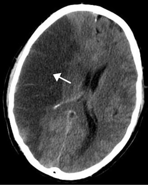 | |
| 脳のCTスキャンでは、動脈閉塞による大規模な右側虚血性脳卒中が以前に起こったことが示されています。CTでは変化が初期には見えない場合があります。 [1] | |
| 専門 | 神経学、脳卒中医学 |
| 症状 | 体の片側が動かない、または感覚がない、理解や会話に問題、めまい、片側の視力喪失[2] [3] |
| 合併症 | 植物状態[4] |
| 原因 | 虚血性(閉塞)と出血性(出血)[5] |
| リスク要因 | 年齢、[6]、 高血圧、喫煙、肥満、高血中コレステロール、糖尿病、一過性脳虚血発作の既往、末期腎不全、心房細動[ 2] [7] [8] |
| 診断方法 | 出血を除外するために通常使用される画像診断による症状に基づく[9] [10] |
| 鑑別診断 | 低血糖[9] |
| 処理 | タイプに基づいて[2] |
| 予後 | 平均寿命1年[2] |
| 頻度 | 4,240万人(2015年)[11] |
| 死亡者(数 | 630万人(2015年)[12] |
脳卒中(脳血管障害(CVA)または脳発作とも呼ばれる)は、脳への血流が悪くなり、脳細胞が死滅する病気です。[5]脳卒中には主に2つの種類があります。
どちらも脳の一部が正常に機能しなくなる原因となります。[5]
脳卒中の兆候や症状には、体の片側が動かなくなったり感じなくなったりすること、理解や会話に問題が生じること、めまい、片側の視力喪失などがあります。[2] [3]兆候や症状は、脳卒中発生後すぐに現れることがよくあります。[3]症状が1~2時間以内に続く場合、その脳卒中は一過性脳虚血発作(TIA)であり、ミニ脳卒中とも呼ばれます。[3] 出血性脳卒中は、激しい頭痛を伴うこともあります。[3]脳卒中の症状は永続的になる可能性があります。[5]長期の合併症には、肺炎や膀胱のコントロールの喪失などがあります。[3]
脳卒中の最大の危険因子は高血圧である。[7]その他の危険因子には、高コレステロール血症、喫煙、肥満、糖尿病、過去の一過性脳虚血発作、末期腎疾患、心房細動などがある。[2] [7] [8]虚血性脳卒中は典型的には血管の閉塞によって引き起こされるが、それほど一般的ではない原因もある。[13] [14] [15]出血性脳卒中は、脳内への直接出血または脳の膜の間への出血によって引き起こされる。[13] [16]出血は、脳動脈瘤の破裂によって起こることがある。[13]診断は通常、身体診察に基づいて行われ、CTスキャンやMRIスキャンなどの医療画像診断によって裏付けられる。[9] CTスキャンでは出血を除外できるが、初期にはCTスキャンでは通常表示されない虚血を必ずしも除外できるわけではない。[10]心電図(ECG)や血液検査などの他の検査は、危険因子を特定し、他の考えられる原因を除外するために行われます。[9]低血糖も同様の症状を引き起こす可能性があります。[9]
予防には、危険因子の減少、頸動脈狭窄が問題となっている人に対する脳への動脈を開く手術、心房細動の人に対するワルファリンの投与などがある。[2]予防のために医師がアスピリンやスタチンを推奨することもある。[2]脳卒中は医学的な緊急事態である。[5]虚血性脳卒中は、3~4時間半以内に発見されれば、血栓を分解する薬で治療できる可能性があるが、[2]出血性脳卒中は手術が有効な場合がある。[2]失われた機能の回復を試みる治療は脳卒中リハビリテーションと呼ばれ、理想的には脳卒中ユニットで行われるが、世界の多くの地域ではこのような施設は利用できない。[2]
2023年には、世界中で1500万人が脳卒中を患った。[17] 2021年には、脳卒中は死亡原因の第3位となり、全死亡数の約10%を占めた。[18] 2015年には、脳卒中を患いながらも生存している人は約4240万人であった。[11] 1990年から2010年の間に、先進国では脳卒中の年間発生率が約10%減少したが、発展途上国では10%増加した。[19] 2015年には、脳卒中は冠動脈疾患に次いで2番目に多い死因となり、630万人の死者(全体の11%)を占めた。[12]約300万人が虚血性脳卒中で死亡し、330万人が出血性脳卒中で死亡した。[12]脳卒中を患った人の約半数は1年未満しか生きられない。[2]全体として、脳卒中の症例の3分の2は65歳以上で発生しました。[19]
分類

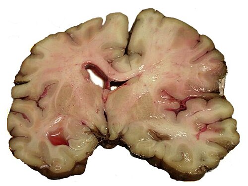
脳卒中は、虚血性と出血性の2つの主要なカテゴリーに分類できます。[20]虚血性脳卒中は脳への血液供給が途絶えることで発生し、出血性脳卒中は血管の破裂または異常な血管構造によって発生します。
脳卒中の約87%は虚血性で、残りは出血性です。虚血部位内で出血が発生することがあり、この状態は「出血性変化」として知られています。出血性脳卒中の何例が実際に虚血性脳卒中として始まるかは不明です。[2]
意味
1970年代に世界保健機関は「脳卒中」を「24時間を超えて持続するか、24時間以内に死亡によって中断される脳血管性神経障害」と定義した[21]が、「脳卒中」という言葉自体が何世紀も前から使われている。この定義は組織損傷の可逆性を反映するはずで、その目的で考案されたもので、24時間という時間枠は恣意的に選択された。24時間という制限は、脳卒中と一過性脳虚血発作を区別するものである。一過性脳虚血発作は、24時間以内に完全に消失する脳卒中症状の関連症候群である。[2]早期に投与すれば脳卒中の重症度を軽減できる治療法が利用できるようになったため、現在では多くの人が「脳卒中」や「急性虚血性脳血管症候群」(それぞれ心臓発作と急性冠症候群をモデルにしている)などの代替用語を好んで使用し、脳卒中症状の緊急性と迅速な対応の必要性を反映している。[22]
虚血性
虚血性脳卒中が発生すると、脳の一部への血液供給が減少し、その領域の脳組織の機能不全を引き起こします。これが起こる理由は 4 つ考えられます。
明らかな原因のない脳卒中は、原因不明の脳卒中(特発性)と呼ばれ、虚血性脳卒中の全症例の30~40%を占めます。[2] [25]
急性虚血性脳卒中には分類システムがある。オックスフォードコミュニティ脳卒中プロジェクト分類(OCSP、バンフォード分類またはオックスフォード分類とも呼ばれる)は主に初期症状に基づいており、症状の程度に基づいて、脳卒中エピソードは全前循環梗塞(TACI)、部分前循環梗塞(PACI)、ラクナ梗塞(LACI)、または後循環梗塞(POCI)に分類される。これら4つの項目は、脳卒中の程度、影響を受ける脳の領域、根本的な原因、および予後を予測する。[26] [27]
TOAST(急性脳卒中治療におけるOrg 10172試験)分類は、臨床症状とさらなる検査結果に基づいており、これに基づいて脳卒中は以下に分類されます。
(1)大動脈の 動脈硬化による血栓症または塞栓症、
(2)心臓に由来する塞栓症、
(3)小血管の完全な閉塞、
(4)その他の決定的な原因
(5)原因不明(原因が2つ考えられる、原因が特定されていない、または調査が不完全)[28]
コカインやメタンフェタミンなどの覚醒剤の使用者は虚血性脳卒中のリスクが高い。[29]
出血性

出血性脳卒中には主に2つのタイプがある: [30] [31]
- 脳内出血は、脳自体の内部で起こる出血(脳内の動脈が破裂し、周囲の組織に血液が溢れる)であり、脳実質内出血(脳組織内での出血)または脳室内出血(脳室系内での出血)が原因です。
- くも膜下出血は、脳組織の外側ではあるものの頭蓋骨内部、正確にはくも膜と軟膜(脳を囲む髄膜の 3 層のうち最も内側の繊細な層)の間で起こる出血です。
上記の2つの主なタイプの出血性脳卒中は、頭蓋内のどこかに血液が蓄積する頭蓋内出血の2つの異なる形態でもあります。しかし、硬膜外血腫(頭蓋骨と硬膜(脳を囲む髄膜の最も外側の厚い層)の間の出血)や硬膜下血腫(硬膜下腔内の出血)などの他の形態の頭蓋内出血は、「出血性脳卒中」とはみなされません。[32]
出血性脳卒中は、脳アミロイド血管症、脳動静脈奇形、頭蓋内動脈瘤などの脳血管の変化を背景に発生する可能性があり、脳実質内出血やくも膜下出血を引き起こす可能性があります。[33]
出血性脳卒中は、神経障害に加えて、通常、特定の症状(例えば、くも膜下出血は典型的には雷鳴頭痛と呼ばれる激しい頭痛を引き起こします)を引き起こしたり、以前の頭部外傷の証拠を明らかにしたりします。
兆候と症状
脳卒中の症状は、通常、数秒から数分で突然始まり、ほとんどの場合それ以上進行しません。症状は脳の影響を受ける領域によって異なります。脳の影響を受ける領域が広いほど、失われる機能が多くなります。脳卒中の一部の形態では、追加の症状が発生する場合があります。たとえば、頭蓋内出血では、影響を受けた領域が他の構造を圧迫する可能性があります。くも膜下出血、脳静脈血栓症、およびまれに脳内出血を除いて、ほとんどの脳卒中は頭痛を伴いません。 [ 33]
早期認識

脳卒中の認識を高めるためのシステムが提案されている。突然の顔面筋力低下、腕のずれ(両腕を上げるように指示されたときに、無意識に片方の腕が下がってしまう)、異常な発話は、脳卒中の症例を正しく特定できる可能性が最も高い所見であり、これらのうち少なくとも 1 つが存在する場合、その可能性は 5.5 倍に増加する。同様に、これら 3 つすべてが存在しない場合、脳卒中の可能性は減少する(尤度比0.39)。[34]これらの所見は脳卒中の診断には最適ではないが、比較的迅速かつ容易に評価できるため、急性期の環境では非常に価値がある。
脳卒中の警告サインを覚えるための記憶法はFAST(顔面下垂、腕の脱力、発話困難、救急サービスを呼ぶまでの時間)[35]であり、保健省(英国)と脳卒中協会、米国脳卒中協会、全米脳卒中協会(米国)によって提唱されている。FASTは後方循環脳卒中の認識においては信頼性が低い。[36]この欠点を補い、脳卒中の早期発見をさらに向上させるために、評価項目にバランス(歩行中または立っているときに突然バランスを保つのが困難になる)と視力(ぼやけた視界や複視の新たな発症、または突然の痛みのない視力喪失)を加えた改訂版記憶法BE FASTが提案されている。[37] [38]脳卒中の病院前検出のための他のスケールには、ロサンゼルス病院前脳卒中スクリーン(LAPSS)[39]とシンシナティ病院前脳卒中スケール(CPSS) [40]があり、FAST法はこれらを基にしている。[41]これらのスケールの使用は専門家のガイドラインで推奨されています。[42]
救急外来に搬送された患者にとって、脳卒中の早期発見は診断検査や治療を迅速化するために重要であると考えられています。この目的のために、病歴と身体検査の特徴に基づいたROSIER(救急外来における脳卒中の認識)と呼ばれるスコアリングシステムが推奨されています。[42] [43]
関連する症状
意識喪失、頭痛、嘔吐は、通常、血栓症よりも出血性脳卒中の方が頻繁に起こります。これは、漏れた血液が脳を圧迫して 頭蓋内圧が上昇するためです。
症状が発症時に最大である場合、原因はくも膜下出血または塞栓性脳卒中である可能性が高くなります。
サブタイプ
影響を受けた脳の領域に、脊髄視床路、皮質脊髄路、および脊髄後柱-内側毛帯路という3 つの主要な中枢神経系の経路のいずれかが含まれる場合、次のような症状が現れることがあります。
ほとんどの場合、症状は体の片側(片側性)にのみ現れます。脳の欠陥は通常、体の反対側にあります。しかし、これらの経路は脊髄にも伝わり、脊髄の損傷もこれらの症状を引き起こす可能性があるため、これらの症状のいずれかが存在することは必ずしも脳卒中を示すものではありません。上記の中枢神経系の経路に加えて、脳幹は12 の脳神経のほとんどを生み出します。したがって、脳幹と脳に影響を与える脳幹脳卒中は、これらの脳神経の欠陥に関連する症状を引き起こす可能性があります。[引用が必要]
- 嗅覚、味覚、聴覚、視覚の変化(全体的または部分的)
- まぶたの垂れ下がり(眼瞼下垂)と眼筋の衰弱
- 反射神経の低下:嘔吐、嚥下、光に対する瞳孔反応
- 顔面の感覚低下および筋力低下
- バランス障害と眼振
- 呼吸と心拍数の変化
- 胸鎖乳突筋の筋力が弱くなり、頭を片側に回すことができない
- 舌の弱さ(舌を突き出したり、左右に動かすことができない)
大脳皮質が影響を受けると、中枢神経系の経路が再び影響を受ける可能性がありますが、次のような症状も現れることがあります。
- 失語症(言語表現、聴覚理解、読み書きの困難、典型的にはブローカ野またはウェルニッケ野が関与)
- 構音障害(神経損傷に起因する運動性言語障害)
- 失行症(随意運動の異常)
- 視野欠損
- 記憶障害(側頭葉の障害)
- 半側無視(頭頂葉の障害)
- 思考の混乱、混乱、性欲過剰の行動(前頭葉の関与を伴う)
- 通常は脳卒中に関連する障害に対する認識の欠如
小脳が関与している場合は、運動失調が存在する可能性があり、これには以下が含まれます。
先行する兆候と症状
脳卒中が起こる数日前(通常は脳卒中が起こる前の7日間、あるいはその直前)に、かなりの割合の患者が「前兆頭痛」を経験します。これは、問題を示す重篤で異常な頭痛です。[45]この症状が現れた場合は、医師の診察を受け、脳卒中の予防を検討することが推奨されます。
原因
血栓性脳卒中

血栓性脳卒中では、通常、アテローム性動脈硬化性プラークの周囲に血栓[46] (血のかたまり) が形成されます。動脈の閉塞は徐々に進行するため、症状のある血栓性脳卒中の発症は出血性脳卒中よりも遅くなります。血栓自体は (血管を完全に塞いでいなくても) 破れて血流に乗って移動すると塞栓性脳卒中 (下記参照) を引き起こす可能性があり、その時点で塞栓と呼ばれます。2種類の血栓症が脳卒中を引き起こす可能性があります。
- 大血管疾患には、総頸動脈、内頸動脈、椎骨動脈、ウィリス動脈輪が関与する。[47]大血管に血栓を形成する可能性のある疾患には、(発生率の高い順に)動脈硬化症、血管収縮(動脈の締め付け)、大動脈解離、頸動脈または椎骨動脈解離、血管壁の炎症性疾患(高安動脈炎、巨細胞性動脈炎、血管炎)、非炎症性血管症、モヤモヤ病、線維筋性異形成などがある。動脈解離によって引き起こされる脳卒中は、厳密な意味では必ずしも「定義された病状」によって引き起こされるわけではなく、そのような事象は非常に若い人々に発生する可能性があり、首の過伸展などの身体的損傷、または多くの場合他の形態の外傷によって引き起こされる可能性がある。[48]
- 小血管疾患は、脳内のより小さな動脈、すなわちウィリス動脈輪の枝、中大脳動脈、動脈幹、および遠位椎骨動脈と脳底動脈から発生する動脈に影響を及ぼす。 [49]小血管に血栓を形成する可能性のある疾患には、(発生率の高い順に)脂肪硝子体症(高血圧と加齢の結果として血管内に脂肪性硝子体が蓄積する)およびフィブリノイド変性(これらの血管が関与する脳卒中はラクナ脳卒中として知られている)、および微小アテローム(小さなアテローム性プラーク)がある。 [50]
貧血は血液循環系における血流増加を引き起こします。これにより血管の内皮細胞が接着因子を発現し、血液凝固と血栓形成を促進します。[51] 鎌状赤血球貧血は血液細胞が凝集して血管を塞ぐ原因となり、脳卒中を引き起こすこともあります。脳卒中は鎌状赤血球貧血の20歳未満の人の死亡原因の第2位です。[52] 大気汚染も脳卒中のリスクを高める可能性があります。[53]
塞栓性脳卒中
塞栓性脳卒中とは、動脈塞栓(動脈の閉塞)が、他の場所から動脈血流中を移動する粒子または破片である塞栓によって起こることを指します。塞栓は血栓であることが最も一般的ですが、脂肪(骨折時の骨髄など)、空気、癌細胞、細菌の塊(通常は感染性心内膜炎による)など、他のさまざまな物質であることもあります。[54]
塞栓は他の場所から発生するため、局所療法では一時的にしか問題を解決できません。したがって、塞栓の発生源を特定する必要があります。塞栓による閉塞は突然発生するため、症状は通常、開始時に最大になります。また、塞栓が部分的に吸収されて別の場所に移動したり、完全に消失したりすると、症状は一時的なものになる場合があります。
塞栓は心臓(特に心房細動)から発生することが最も多いが、動脈樹の他の部位から発生することもある。逆説的塞栓症では、深部静脈血栓が心臓の心房中隔欠損または心室中隔欠損を介して脳に塞栓する。 [54]
心臓に関連する脳卒中の原因は、高リスクと低リスクに分けられます。[55]
- 高リスク:心房細動および発作性心房細動、僧帽弁または大動脈弁のリウマチ性疾患、人工心臓弁、心房または心室の既知の心血栓、洞不全症候群、持続性心房粗動、最近の心筋梗塞、駆出率< 28%を伴う慢性心筋梗塞、駆出率 < 30% を伴う症候性うっ血性心不全、拡張型心筋症、リブマン・サックス心内膜炎、マランティック心内膜炎、感染性心内膜炎、乳頭線維弾性腫、左房粘液腫、および冠動脈バイパス移植(CABG) 手術。
- 低リスク/低潜在性: 僧帽弁輪の石灰化、卵円孔開存 (PFO)、心房中隔瘤、卵円孔開存を伴う心房中隔瘤、血栓のない左室瘤、心エコー検査での左房単独の「煙」(僧帽弁狭窄または心房細動なし)、および上行大動脈または近位弓部の複雑なアテローム
頸動脈の1つが完全に閉塞している人の場合、その側の脳卒中リスクは年間約1%です。[56]
塞栓性脳卒中の特殊な形態として、原因不明塞栓性脳卒中(ESUS)がある。この原因不明脳卒中は、近位動脈狭窄や心臓塞栓性の原因がない非ラクナ性脳梗塞と定義される。虚血性脳卒中の約6例中1例がESUSに分類される可能性がある。[57]
脳低灌流
脳低灌流とは、脳のすべての部分への血流が減少することです。原因によっては、脳の特定の部分で血流が減少する可能性があります。最も一般的な原因は、心停止または不整脈による心不全、または心筋梗塞、肺塞栓症、心嚢液貯留、または出血の結果としての心拍出量の低下です。 [要出典]低酸素症(血液中の酸素含有量が少ない)が低灌流を促進する可能性があります。血流の減少は脳全体に及ぶため、脳のすべての部分が影響を受ける可能性がありますが、特に脆弱な「分水嶺」領域(主要な脳動脈によって血液が供給される境界領域)が影響を受けます。分水嶺脳卒中とは、これらの領域への血液供給が損なわれた状態を指します。これらの領域への血流は必ずしも停止するわけではありませんが、脳損傷が発生する可能性があるレベルまで減少する可能性があります。
静脈血栓症
脳静脈洞血栓症は、局所的に静脈圧が上昇し、動脈によって生成される圧力を超えることで脳卒中を引き起こします。梗塞は、他のタイプの虚血性脳卒中よりも出血性変化(損傷領域への血液の漏出)を起こす可能性が高くなります。[24]
脳内出血
一般的には小動脈または細動脈に発生し、高血圧、[58]頭蓋内血管奇形(海綿状血管腫または動静脈奇形を含む)、脳アミロイド血管症、または二次出血が起こった梗塞が原因であることが多い。[2]その他の潜在的な原因としては、外傷、出血性疾患、アミロイド血管症、違法薬物使用(例:アンフェタミンまたはコカイン)がある。血腫は、周囲の組織からの圧力によって成長が制限されるまで、または脳室系、脳脊髄液、または軟膜表面に排出されて減圧されるまで拡大する。脳内出血の3分の1は脳室で起こる。 ICHの死亡率は30日後に44%となり、虚血性脳卒中やくも膜下出血(技術的には脳卒中の一種としても分類される可能性がある[2]) よりも高い。
他の
その他の原因としては、動脈のけいれんが挙げられます。これはコカインによって起こることがあります。[59] がんもまた、脳卒中の潜在的原因としてよく知られています。悪性腫瘍は一般的に脳卒中のリスクを高めますが、膵臓がん、肺がん、胃がんなどの特定の種類のがんは、通常、血栓塞栓症のリスクが高くなります。がんが脳卒中のリスクを高めるメカニズムは、後天的な凝固亢進に続発すると考えられています。[60]
サイレントストローク
無症候性脳卒中は外見上の症状がなく、本人は通常、脳卒中を経験したことに気づいていない。識別可能な症状を引き起こさないにもかかわらず、無症候性脳卒中は脳に損傷を与え、将来的に一過性脳虚血発作と重度脳卒中の両方のリスクが増加する。逆に、重度脳卒中を経験した人は無症候性脳卒中になるリスクもある。 [61] 1998年の広範な調査では、米国では1100万人以上が脳卒中を経験したと推定されている。このうち約77万人が症候性で、1100万人が初めてのMRIによる無症候性梗塞または出血だった。無症候性脳卒中では通常、MRIなどの神経画像診断によって検出される病変を引き起こす。無症候性脳卒中は症候性脳卒中の5倍の割合で発生すると推定されている。[62] [63]無症候性脳卒中のリスクは加齢とともに増加するが、特に急性貧血の若年成人や小児にも影響を及ぼす可能性がある。[62] [64]
病態生理学
虚血性

虚血性脳卒中は、脳の一部への血液供給が失われ、虚血カスケードが開始されるために発生します。[65]アテローム性動脈硬化症は、血管の内腔を狭めて血液供給を妨げ、血管内に血栓を形成して血流を減少させるか、アテローム性動脈硬化性プラークの崩壊によって小さな塞栓が大量に放出されることがあります。[66]塞栓性梗塞は、循環器系の他の部分、典型的には心房細動の結果として心臓内、または頸動脈内で形成された塞栓が剥がれて脳循環に入り、脳血管に詰まって閉塞するときに発生します。脳の血管が閉塞しているため、脳はエネルギー不足になり、虚血の影響を受けた脳組織領域内で嫌気性代謝を使用するようになります。嫌気性代謝ではアデノシン三リン酸(ATP)の生成量は少なくなりますが、乳酸と呼ばれる副産物が放出されます。乳酸は酸であるため、脳内の正常な酸塩基バランスを崩し、細胞を破壊する可能性のある刺激物です。虚血領域は「虚血性周辺部」と呼ばれます。[67]最初の虚血イベントの後、周辺部は損傷を特徴とする組織リモデリングから修復を特徴とするリモデリングに移行します。[68]

虚血性脳組織で酸素やグルコースが枯渇すると、アデノシン三リン酸(ATP)などの高エネルギーリン酸化合物の生成が失敗し、組織細胞の生存に必要なエネルギー依存プロセス(イオンポンプなど)が機能しなくなります。これにより、一連の相互に関連したイベントが発生し、細胞の損傷と死につながります。ニューロン損傷の主な原因は、興奮性神経伝達物質グルタミン酸の放出です。神経系の細胞外のグルタミン酸濃度は通常、細胞膜を横切るイオン(主にNa +)の濃度勾配によって駆動される、いわゆる取り込みキャリアによって低く抑えられています。しかし、脳卒中により、これらの勾配を維持するイオンポンプの駆動源である酸素とグルコースの供給が遮断されます。その結果、膜貫通イオン勾配が低下し、グルタミン酸トランスポーターが方向を逆転して、グルタミン酸を細胞外空間に放出します。グルタミン酸は神経細胞の受容体(特にNMDA受容体)に作用し、細胞のタンパク質、脂質、核物質を消化する酵素を活性化するカルシウムの流入を引き起こします。カルシウムの流入はミトコンドリアの機能不全にもつながり、それがエネルギー枯渇につながり、プログラム細胞死による細胞死を引き起こす可能性があります。[69]
虚血は、酸素フリーラジカルやその他の活性酸素種の生成も引き起こします。これらは、多くの細胞および細胞外要素と反応して損傷を与えます。血管の内層や内皮が損傷を受ける可能性があります。これらのプロセスは、どのタイプの虚血組織でも同じであり、総称して虚血カスケードと呼ばれます。ただし、脳組織は、他のほとんどの臓器とは異なり、呼吸予備力がほとんどなく、好気性代謝に完全に依存しているため、特に虚血に対して脆弱です。
担保フロー
_Schematic_drawing_of_the_collateral_network_showing_anastomoses_between_the_middle_cerebral_artery_(MCA)_and_anterior_cerebral_artery.png/500px-thumbnail.png)
脳は、単一の動脈の血流不足を側副血行路によって補うことができる。このシステムは、ウィリス動脈輪と、それほどではないが大脳半球に血液を供給する主要動脈を介した頸動脈と椎骨動脈の効率的な接続に依存している。しかし、ウィリス動脈輪の変化、側副血管の口径、および動脈硬化などの後天性動脈病変は、この補償メカニズムを妨害し、動脈閉塞による脳虚血のリスクを高める可能性がある。[70]
損傷の程度は、虚血の持続時間と重症度によって異なります。灌流が正常の 5% 未満で虚血が 5 分以上続くと、一部のニューロンが死滅します。ただし、虚血が軽度の場合、損傷はゆっくりと進行し、脳組織が完全に破壊されるまでに最大 6 時間かかることがあります。重度の虚血が 15 ~ 30 分以上続くと、影響を受けた組織がすべて死滅し、梗塞につながります。損傷の速度は温度の影響を受け、高体温は損傷を加速し、低体温は損傷を遅らせるなど、他の要因があります。虚血組織への血流を速やかに回復させることで、特に組織がまだ不可逆的な損傷を受けていない場合は、損傷を軽減または回復させることができます。これは、重度の虚血領域の周囲の中等度の虚血領域 (周辺部) にとって特に重要であり、側副血行によりまだ救済できる可能性があります。[70] [71] [72]
出血性
出血性脳卒中は、その基礎病理に基づいて分類される。出血性脳卒中の原因には、高血圧性出血、動脈瘤破裂、房室瘻破裂、以前の虚血性梗塞の変化、薬剤誘発性出血などがある。[73]これらは、拡大する血腫による組織の圧迫を引き起こし、組織損傷を引き起こす。さらに、圧力により影響を受けた組織への血液供給が失われ、結果として梗塞が発生する可能性があり、脳出血によって放出された血液は脳組織と血管に直接的な毒性作用を及ぼすと思われる。[52] [74] 炎症は出血後の二次的脳損傷に寄与する。 [74]
診断


脳卒中は、神経学的検査( NIHSSなど)、CTスキャン(ほとんどの場合、造影剤を使用しない)またはMRIスキャン、ドップラー超音波、動脈造影など、いくつかの技術で診断されます。脳卒中自体の診断は、画像診断技術の助けを借りて臨床的に行われます。画像診断技術は、脳卒中のサブタイプと原因の特定にも役立ちます。脳卒中の診断自体に一般的に使用される血液検査はまだありませんが、血液検査は脳卒中の考えられる原因を見つけるのに役立つ場合があります。[75]死亡した人の場合、脳卒中の剖検は、脳卒中の発症から死亡までの時間を確定するのに役立つ場合があります。
身体検査
身体検査では、症状の病歴や神経学的状態を調べることで、脳卒中が発生した場所や重症度を評価することができます。身体検査では、例えばNIH 脳卒中スケールの標準スコアを出すことができます。
イメージング
緊急時の虚血性脳卒中の診断:[76]
緊急時の出血性脳卒中の診断の場合:
- CTスキャン(造影剤
なし)
- 感度= 89%
- 特異度 = 100%
- MRI スキャン
- 感度= 81%
- 特異度 = 100%
慢性出血の検出にはMRIスキャンの方が感度が高い。[77]
安定した脳卒中の評価には、単一光子放出コンピュータ断層撮影(SPECT) や陽電子放出断層撮影-コンピュータ断層撮影(PET/CT) などの核医学スキャンが役立つ場合があります。SPECT は脳血流を記録しますが、FDG 同位体を使用した PET は脳のグルコース代謝を示します。
CTスキャンでは、特に脳卒中が小さく、発症して間もない場合[10]や、脳幹や小脳領域(後循環梗塞)の場合、虚血性脳卒中を検出できないことがあります。MRIは拡散強調画像で後循環梗塞を検出するのにより適しています。[78] CTスキャンは、特定の脳卒中類似症状を除外し、出血を検出するために使用されます。 [10]脳内の軟膜側副循環の存在は、再開通治療後のより良い臨床結果と関連しています。 [79]脳血管予備能は、脳卒中の結果に影響を与えるもう1つの要因です。これは、吸入二酸化炭素や静脈内 アセタゾラミドの投与など、医師による意図的な血流刺激後の脳血流量の増加量です。血流量の増加は、PETスキャンまたは経頭蓋ドップラー超音波検査で測定できます。[80]しかし、片側の内頸動脈が閉塞している人では、軟膜側副循環の存在が脳予備能の低下と関連している。[81]
根本的な原因
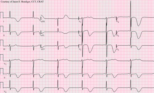
脳卒中と診断された場合、根本的な原因を突き止めるために他の検査が行われることがあります。利用可能な治療と診断のオプションを考慮すると、末梢に塞栓源があるかどうかを判断することが特に重要です。脳卒中の原因は年齢、併存疾患、臨床症状によって異なるため、検査の選択は異なる場合があります。一般的に使用される技術は次のとおりです。
- 頸動脈の超音波/ドップラー検査(頸動脈狭窄を検出するため)または脳前動脈の解離。
- 心電図( ECG)と心エコー図(血流を通じて脳血管に広がる可能性がある心臓内の不整脈とその結果生じる血栓を特定するため)
- 断続的な異常な心拍リズムを特定するためのホルター心電図検査。
- 脳血管の血管造影検査(出血が動脈瘤または動静脈奇形から発生したと考えられる場合)
- 血液検査により、血中コレステロール値が高いかどうか、異常な出血傾向があるかどうか、ホモシスチン尿症などのまれな病気が関与しているかどうかを確認します。
出血性脳卒中の場合、血管内造影剤を用いたCTまたはMRIスキャンにより、脳動脈の異常(動脈瘤など)やその他の出血源を特定できる可能性があり、原因が見つからない場合は構造的MRIが行われます。それでも出血の根本的な原因が特定されない場合は、侵襲性脳血管造影を行うこともできますが、血管内カテーテルで血流にアクセスする必要があり、挿入部位の合併症だけでなくさらなる脳卒中を引き起こす可能性があるため、この検査は特定の状況にのみ行われます。[82]出血が静脈血栓症の結果として起こった可能性があることを示唆する症状がある場合は、CTまたはMRI静脈造影を使用して脳静脈を検査できます。[82]
誤診
虚血性脳卒中患者のうち、誤診は2~26%の割合で起こる。[83]「脳卒中カメレオン」(SC)とは、別の病気として診断される脳卒中のことである。[83] [84]
脳卒中を患っていない人も、脳卒中と誤診されることがある。このような場合に血栓溶解剤(血栓を溶かす薬)を投与すると、脳内出血が 1 ~ 2% の確率で起こるが、これは脳卒中患者の場合よりも少ない。この不必要な治療は医療費を増大させる。それでも、AHA/ASA ガイドラインでは、類似症状の可能性がある場合には、追加検査のために治療を遅らせるよりも、tPA の静脈内投与を開始することが望ましいとされている。[83]
女性、アフリカ系アメリカ人、ヒスパニック系アメリカ人、アジア系および太平洋諸島系は、実際には脳卒中であるにもかかわらず、脳卒中以外の病状と誤診されることが多い。さらに、44歳未満の成人は、75歳以上の成人よりも脳卒中を見逃す可能性が7倍高い。これは、特に後循環梗塞のある若い人に当てはまる。[83]一部の医療センターでは、当初脳卒中の可能性が低いと考えられていた人々を対象に、実験研究で超急性MRIを使用しており、これらの人々の一部に脳卒中が見つかり、血栓溶解薬で治療された。[83]
防止
脳卒中の疾病負担を考えると、予防は重要な公衆衛生上の懸念事項である。[85 ] 一次予防は二次予防よりも効果が低い(1年間に1件の脳卒中を予防するために必要な治療件数で判断)。 [85]最近のガイドラインでは、脳卒中の一次予防に関するエビデンスが詳述されている。[86]脳卒中の予防薬としてのアスピリンの使用については、健康な人ではアスピリンは有益ではないようで推奨されないが、[87] 心血管リスクが高い人や心筋梗塞を起こしたことがある人では、初回脳卒中に対してある程度の予防効果がある。[88] [89]以前に脳卒中を起こしたことがある人では、アスピリン、クロピドグレル、ジピリダモールなどの薬剤による治療が有益な場合がある。[88]米国予防医学タスクフォース(USPSTF)は、症状のない人に対して頸動脈狭窄のスクリーニングを行わないよう推奨している。[90]
リスク要因
脳卒中の最も重要な修正可能な危険因子は高血圧と心房細動ですが、その影響は小さく、1回の脳卒中を予防するためには833人を1年間治療する必要があります。[91] [92]その他の修正可能な危険因子には、高コレステロール血症、糖尿病、末期腎疾患、[8]喫煙[93] [94](能動的および受動的)、多量のアルコール摂取、[95]薬物使用、[96]身体活動不足、肥満、加工赤肉の摂取、[97]および不健康な食事[98]などがあります。1日1本のタバコを吸うだけでも、リスクは30%以上増加します。[99]アルコール摂取は、複数のメカニズム(例えば、高血圧、心房細動、反跳血小板増多症、血小板凝集および凝固障害)を介して、虚血性脳卒中、ならびに脳内出血およびくも膜下出血を起こしやすくなる可能性がある。 [100]薬物、最も一般的なのはアンフェタミンとコカインであり、脳の血管への損傷および急性高血圧を介して脳卒中を引き起こす可能性がある。[73] [101] 前兆を伴う片頭痛は、虚血性脳卒中のリスクを2倍にする。[102] [103]症状の有無にかかわらず、治療されないセリアック病は、子供と大人の両方で脳卒中の根本的な原因となり得る。[104] 2021年のWHOの調査によると、週55時間以上働くと、週35~40時間働く場合と比較して、脳卒中のリスクが35%、心臓病で死亡するリスクが17%上昇します。[105]
高いレベルの身体活動は脳卒中のリスクを約26%減少させます。[106]生活習慣の改善を促す取り組みを調べた質の高い研究は不足しています。[107]それでも、状況証拠が多数あることを考慮すると、脳卒中の最善の医学的管理には、食事、運動、喫煙、飲酒に関するアドバイスが含まれます。[108]薬物療法は脳卒中予防の最も一般的な方法ですが、頸動脈内膜剥離術は脳卒中を予防する有用な外科的治療法となり得ます。
血圧
高血圧は脳卒中リスクの35~50%を占める。[109]収縮期血圧を10 mmHg、拡張期血圧を5 mmHg下げると、脳卒中リスクが約40%低下する。[110]血圧を下げることは、虚血性脳卒中と出血性脳卒中の両方を予防することが決定的に示されている。[111] [112]二次予防においても同様に重要である。[113] 80歳以上の人や収縮期高血圧のみの人でも、降圧療法の恩恵を受けることができる。[114] [115] [116]利用可能なエビデンスでは、降圧薬による脳卒中予防に大きな違いは見られないため、他の心血管疾患の予防やコストなどの他の要因も考慮する必要がある。[117] [118]脳卒中または一過性脳虚血発作後のβ遮断薬の日常的な使用が有益であることは示されていない。[119]
血中脂質
高コレステロール値は(虚血性)脳卒中と一貫して関連していない。[112] [120] スタチンは脳卒中のリスクを約15%低下させることが示されている。[121]他の脂質低下薬の以前のメタアナリシスではリスクの低下が示されなかったため、[122]スタチンは脂質低下効果以外のメカニズムを通じて効果を発揮する可能性がある。[121]
糖尿病
糖尿病は脳卒中のリスクを2~3倍に高めます。[要説明] [要出典]徹底的な血糖コントロールは腎臓障害や目の網膜損傷などの小血管合併症を軽減することが示されていますが、脳卒中などの大血管合併症を軽減することは示されていません。[123] [124]
抗凝固薬
ワルファリンなどの経口抗凝固薬は、50年以上にわたって脳卒中予防の主流となってきました。しかし、いくつかの研究では、アスピリンやその他の抗血小板薬が脳卒中や一過性脳虚血発作後の二次予防に非常に効果的であることが示されています。 [88]低用量のアスピリン(たとえば75~150 mg)は高用量と同等の効果がありますが、副作用が少なく、最小有効用量は不明です。[125] チエノピリジン(クロピドグレル、チクロピジン)は、アスピリンよりもわずかに効果が高く、消化管出血のリスクが低い可能性がありますが、より高価です。[126]アスピリンとクロピドグレルはどちらも、軽度の脳卒中または高リスクTIA後の最初の数週間に有効である可能性があります。[127]クロピドグレルはチクロピジンよりも副作用が少ないです。[126] 頭痛は一般的な副作用ですが、ジピリダモールをアスピリン療法に追加して、わずかな追加効果を得ることができます。 [128]低用量アスピリンは心筋梗塞後の脳卒中予防にも効果的である。[89]
心房細動の患者は脳卒中のリスクが年間5%あり、弁膜性心房細動の患者はさらに高いリスクがあります。[129]脳卒中のリスクに応じて、ワルファリンやアスピリンなどの薬剤による抗凝固療法は予防に有用であり、使用される治療の種類に応じて比較有効性のレベルは異なります。 [130] [131]
経口抗凝固薬、特にXa(アピキサバン)およびトロンビン(ダビガトラン)阻害剤は、脳卒中の軽減においてワルファリンより優れていることが示されており、心房細動患者における出血リスクはワルファリンより低いか同等である。[131]心房細動患者を除いて、経口抗凝固薬は脳卒中予防には推奨されていない。いかなる利点も出血リスクによって相殺されるからである。[132]
しかし、一次予防では抗血小板薬は虚血性脳卒中のリスクを低下させず、大出血のリスクを増加させた。[133] [134]女性における虚血性脳卒中に対するアスピリンの予防効果の可能性を調査するには、さらなる研究が必要である。[135] [136]
手術
頸動脈内膜剥離術または頸動脈形成術は、頸動脈のアテローム性動脈硬化による狭窄を除去するために使用できます。特定の症例では、この手順を支持する証拠があります。[108]有意狭窄に対する頸動脈内膜剥離術は、すでに脳卒中を患っている人のさらなる脳卒中を予防するのに有効であることが示されています。 [137]頸動脈ステント留置術は、同様に有効であるとは示されていません。 [138] [139]手術の対象となる人は、年齢、性別、狭窄の程度、症状出現からの時間、および本人の希望に基づいて選択されます。[108]手術は、あまり長く延期しない場合に最も効果的です。50%以上の狭窄がある人の再発性脳卒中リスクは、5年後に最大20%ですが、頸動脈内膜剥離術によりこのリスクは約5%に低下します。 1人の患者を治すために必要な手術回数は、早期手術(最初の脳卒中後2週間以内)の場合は5回であったが、12週間以上延期した場合は125回であった。[140] [141]
頸動脈狭窄のスクリーニングは、一般集団では有用な検査ではないことが示されています。[142]症状のない頸動脈狭窄に対する外科的介入の研究では、脳卒中のリスクがわずかに低下しただけです。[143] [144]手術が有益であるためには、手術の合併症率を4%未満に抑える必要があります。それでも、100回の手術で、5人が脳卒中を回避して恩恵を受け、3人が手術にもかかわらず脳卒中を発症し、3人が手術自体が原因で脳卒中を発症または死亡し、89人は介入がなかったとしても脳卒中を発症しません。[108]
ダイエット
栄養、特に地中海式の食事は、脳卒中のリスクを半分以上減らす可能性があります。[145]葉酸でホモシステイン値を下げても脳卒中のリスクには影響しないようです。 [146] [147]
女性
女性に対しては、過去に慢性高血圧の病歴がある場合は妊娠11週目以降にアスピリンを服用することや、収縮期血圧が150mmHg以上または拡張期血圧が100mmHg以上の場合は妊娠中に血圧降下剤を服用することなど、いくつかの具体的な推奨事項が出されている。以前に子癇前症を患ったことがある人では、他のリスク要因をより積極的に治療する必要がある。[148]
脳卒中または一過性脳虚血発作の既往
血圧を140/90 mmHg未満に保つことが推奨される。[149]抗凝固療法は再発性虚血性脳卒中を予防することができる。非弁膜症性心房細動の患者の場合、抗凝固療法は脳卒中を60%減らすことができ、抗血小板薬は脳卒中を20%減らすことができる。[150]しかし、最近のメタアナリシスは、抗凝固療法による害は塞栓性脳卒中の直後から始まることを示唆している。[151] [152]心房細動の脳卒中予防治療は、 CHA2DS2-VAScスコアに従って決定される。非弁膜症性心房細動の患者の血栓塞栓性脳卒中を予防するために最も広く使用されている抗凝固薬は経口薬のワルファリンであるが、ダビガトランを含むいくつかの新しい薬剤はプロトロンビン時間のモニタリングを必要としない代替薬である。[149]
脳卒中後に抗凝固薬を使用する場合は、歯科処置のために服用を中止してはならない。[153]
検査で頸動脈狭窄が判明し、患側にある程度の残存機能がある場合、脳卒中後速やかに頸動脈内膜剥離術(狭窄部の外科的除去)を実施することで再発のリスクを軽減できる可能性があります。
管理
脳卒中は、虚血性であれ出血性であれ、直ちに医師の診察を受ける必要がある緊急事態です。[5] [154]具体的な治療法は、脳卒中の種類、症状の発症からの経過時間、根本的な原因や合併症の有無によって異なります。[154]
虚血性脳卒中
アスピリンは再発の全体的なリスクを13%減少させ、早期により大きな利益をもたらします。[155]最初の数時間以内の根治的治療は、血栓を分解する(血栓溶解)か、機械的に除去する(血栓除去)ことによって閉塞を取り除くことを目的としています。迅速な脳卒中介入の重要性の根底にある哲学的前提は、1990年代初頭に「時間は脳だ!」と要約されました。[156]数年後、脳血流の急速な回復により脳細胞の死が減少するという同じ考えが証明され、定量化されました。[157]
最初の数時間の厳格な血糖コントロールは転帰を改善せず、むしろ害を及ぼす可能性がある。[158]高血圧も通常は下げられないが、これは有益であることが分かっていないからである。[159] [160]中国、東欧、ロシア、旧ソ連諸国、その他のアジア諸国で急性虚血性脳卒中の治療に広く使用されている豚脳由来 神経栄養因子の混合物であるセレブロリジンは、転帰を改善せず、死亡を予防せず、重篤な有害事象のリスクを高める可能性がある。 [161 ]牛の脳から抽出されるセレブロリジン様ペプチド混合物が急性虚血性脳卒中の治療に役立つという証拠もない。[161]
血栓溶解
急性虚血性脳卒中における、遺伝子組換え組織プラスミノーゲン活性化因子(rtPA)などによる血栓溶解療法は、症状発現後3時間以内に投与された場合、障害なく生活する上で全体的に10%の利益をもたらす。 [162] [163]しかし、生存の可能性は改善しない。[162]使用が早いほど利益は大きい。[162] 3時間から4時間半の間は、効果は明らかではない。[164] [165] [166] AHA/ASAは、この時間枠内の特定の人々にこれを推奨している。[167] 2014年のレビューでは、3か月から6か月で障害なく生活する人の数が5%増加したが、短期的には死亡リスクが2%増加した。[163] 4時間半を超えると、血栓溶解療法は転帰を悪化させる。[164]これらの利益または利益の欠如は、治療を受けた人の年齢に関係なく発生した。[168]治療後に頭蓋内出血を起こす人と起こさない人を確実に判断する方法はありません。[169] 4.5時間から9時間の間に医療画像で保存可能な組織が見つかった人や脳卒中で目覚めた人には、アルテプラーゼがいくらかの利益をもたらします。[170]
アメリカ心臓協会、アメリカ救急医療学会、アメリカ神経学会は、他の禁忌(異常な臨床検査値、高血圧、最近の手術など)がない限り、症状発現後3時間以内の急性脳卒中の推奨治療としてtPAの使用を承認している。tPAのこの立場は、1つの研究者グループによる2つの研究の結果に基づいており[171]、 tPAは良好な神経学的転帰の可能性を高めることが示された。最初の3時間以内に投与すると、血栓溶解療法は死亡率に影響を与えることなく機能的転帰を改善する。[172]大規模な脳卒中を起こした人の6.4%は、tPA投与による合併症としてかなりの脳出血を起こし、これが短期死亡率の上昇の一因となっている。[173]アメリカ救急医学会は以前、急性虚血性脳卒中に対するtPAの適用性に関する客観的証拠は不十分であると述べていた。[174] 2013年にアメリカ救急医学会はこの立場に反論し、[175]虚血性脳卒中におけるtPAの使用に関する証拠を認めたが、[176]議論は続いている。[177] [178]動脈を通って脳内にカテーテルを挿入し、血栓部位に薬剤を注入する 動脈内線溶療法は、急性虚血性脳卒中の患者の転帰を改善することがわかっている。[179]
血管内治療
虚血性脳卒中の原因となる血栓を機械的に除去する機械的血栓除去術は、中大脳動脈などの大動脈閉塞症の治療法として有望である。2015年に発表されたあるレビューでは、症状の発症から12時間以内にこの処置を実施した場合の安全性と有効性が実証された。[180] [181]機械的血栓除去術の対象となる人に一般的に使用される静脈内血栓溶解療法と比較して、死亡リスクは変わらなかったが、障害は軽減した。[182] [183] 症状の発症から24時間以内に血栓除去術を行うことで、効果が得られる症例もある。[184]
頭蓋骨切除術
脳卒中が脳の大部分に影響を及ぼすと、脳の腫れがひどくなり、周囲の組織に二次的な脳損傷が生じる可能性がある。この現象は主に、中大脳動脈に血液供給を依存する脳組織に影響を及ぼす脳卒中で発生し、予後が不良であることから「悪性脳梗塞」とも呼ばれる。薬物療法で圧力を軽減することも可能だが、片側の頭蓋骨を一時的に外科的に除去する半頭蓋切除術が必要となる場合もある。これにより死亡リスクは減少するが、そうでなければ死亡していたであろう人の一部は障害を負って生き延びる。[185] [186]
出血性脳卒中
脳内出血の患者には、必要に応じて血圧管理を含む支持療法が必要である。意識レベルの変化がないか患者を監視し、血糖値と酸素化を最適なレベルに維持する。抗凝固薬と抗血栓薬は出血を悪化させることがあるため、通常は中止する(可能であれば中止する)。[要出典]一定の割合で、血液を除去して根本原因を治療する脳神経外科的介入が有益となる可能性があるが、これは出血の場所と大きさ、および患者関連の要因に依存しており、どのような脳内出血患者が恩恵を受ける可能性があるかという問題については、現在も研究が行われている。[187]
くも膜下出血では、基礎にある脳動脈瘤を早期に治療することで、さらなる出血のリスクを軽減できる可能性があります。動脈瘤の場所に応じて、頭蓋骨を開く手術や血管内(血管を介した)手術が行われる場合があります。[188]
ストローク単位
理想的には、脳卒中を患った人は、脳卒中治療の経験がある看護師やセラピストが配置された病院内の病棟または専用エリアである「脳卒中ユニット」に入院する。脳卒中ユニットに入院した人は、たとえ脳卒中経験のない医師の治療を受けていても、病院の他の場所に入院した人よりも生存する可能性が高いことがわかっている。[2] [189] 看護ケアは、スキンケア、食事、水分補給、体位の維持、体温、脈拍、血圧などのバイタルサインの監視に不可欠である。 [190]
リハビリテーション
脳卒中リハビリテーションは、日常生活のスキルを回復し再学習することで、障害となる脳卒中患者が可能な限り通常の生活に戻れるように治療を受けるプロセスです。また、生存者が困難を理解して適応できるように支援し、二次的な合併症を防ぎ、家族がサポート役を担えるように教育することも目的としています。脳卒中リハビリテーションは、多分野にわたるアプローチでほぼ直ちに開始する必要があります。リハビリテーション チームには、リハビリテーション医学の訓練を受けた医師、神経科医、臨床薬剤師、看護スタッフ、理学療法士、作業療法士、言語聴覚士、および整形外科医が含まれます。罹患者の少なくとも 3 分の 1 が脳卒中後うつ病を発症するため、一部のチームには心理学者やソーシャルワーカーも含まれる場合があります。Barthelスケールなどの検証済みのツールは、脳卒中を患った人が病院から退院した後、サポートの有無にかかわらず自宅で生活できる可能性を評価するために使用できます。[191]
脳卒中のリハビリテーションはできるだけ早く開始する必要があり、数日から 1 年以上かかる場合があります。ほとんどの機能回復は最初の数か月で見られますが、その後改善は衰え、米国の州のリハビリテーション ユニットやその他の機関によって公式に「ウィンドウ」と見なされる 6 か月後には閉じられ、それ以上改善する可能性はほとんどありません。[医学的引用が必要]ただし、何年も改善を続け、書く、歩く、走る、話すなどの能力を取り戻し、強化したと報告する人もいます。[医学的引用が必要]脳卒中を患った人は、毎日のリハビリテーション運動を日常生活の一部にし続ける必要があります。完全に回復することはまれですが、不可能ではなく、ほとんどの人はある程度改善します。適切な食事と運動は脳の回復を助けることが知られています。
空間無視
認知リハビリテーションが無視による障害を軽減する効果については、十分な証拠が得られておらず、自立性の向上についても証明されていない。[192]しかし、認知リハビリテーションが無視の検査に即時の有益な効果をもたらす可能性があるという証拠は限られている。[192]全体として、空間無視の証拠によって裏付けられるリハビリテーションのアプローチはない。
自動車の運転
リハビリテーションの利用が脳卒中後の路上運転技能を改善できるかどうかは、証拠の集積では不明である。[193]運転シミュレーターでの訓練が訓練後に道路標識の認識能力を向上させるという証拠は限られている。[193]この研究結果は質の低い証拠に基づいており、多数の参加者を対象としたさらなる研究が必要である。
ヨガ
質の低いエビデンスに基づくと、ヨガが生活の質、バランス、筋力、持久力、痛み、障害スコアの指標において脳卒中リハビリテーションに有意な利益をもたらすかどうかは不明である。[194]ヨガは不安を軽減する可能性があり、患者中心の脳卒中リハビリテーションの一部として組み込むことができる。[194]脳卒中リハビリテーションにおけるヨガの利益と安全性を評価するためのさらなる研究が必要である。
上肢の動作観察理学療法
質の低いエビデンスでは、行動観察(ミラーニューロンシステムを通じて神経可塑性を改善することを目的とした理学療法の一種)にはある程度の利益があり、重大な副作用はないが、この利益は臨床的に重要ではない可能性があり、さらなる研究が提案されている。[195]
注意欠陥に対する認知リハビリテーション
脳卒中後の患者の注意欠陥に対する認知リハビリテーションの有効性については、科学的証拠が不十分である。[196]治療後すぐに注意力に効果があるかもしれないが、その知見は質の低い、または中程度の研究と少数の研究に基づいている。[196]注意力を必要とする日常的な作業で効果が持続するかどうかを評価するには、さらなる研究が必要である。
歩行リハビリテーションのための運動イメージ
最新のエビデンスは、他の治療法と比較して、脳卒中を患った人の歩行速度に対する運動イメージ療法(MI)の短期的な利点を支持している。[197] MIは脳卒中後の運動機能を改善せず、重大な有害事象を引き起こすことはないと思われる。[197] MIが歩行持久力と介助への依存度に及ぼす影響を推定するにはさらなる研究が必要であるため、この研究結果は質の低いエビデンスに基づいている。
理学療法と作業療法
理学療法と作業療法には重複した専門分野があるが、理学療法は、運動を実施し、ベッドでの移動、移乗、歩行、その他の粗大運動機能などの機能的タスクを再学習することにより、関節可動域と強度に焦点を当てている。理学療法士は、脳卒中を患った人と協力して、片麻痺側の認識と使用を改善することもできます。リハビリテーションでは、力強い動きを生み出す能力や、正常なパターンを使用してタスクを実行する能力に取り組むことが含まれます。重点は、多くの場合、機能的タスクと患者の目標に集中します。理学療法士が運動学習を促進するために使用する1つの例は、制約誘導運動療法です。継続的な練習により、患者は機能的活動中に片麻痺の手足を使用および適応させることを再学習し、永続的な変化を生み出します。[198]理学療法は、脳卒中後の機能と運動性の回復に効果的です。[199]作業療法は、食べる、飲む、着替える、入浴、調理、読み書き、トイレなどの日常生活動作(ADL)と呼ばれる日常の活動を再学習するためのトレーニングに関係しています。尿失禁患者を助けるためのアプローチには、理学療法、認知療法、経験豊富な医療専門家による専門的な介入などがありますが、これらのアプローチが脳卒中後の尿失禁の改善にどれほど効果的であるかは明らかではありません。[200]
脳卒中に関連する痙縮の治療には、多くの場合、早期の運動療法(一般的には理学療法士が実施)と、痙縮した筋肉の伸長およびさまざまな姿勢での持続的なストレッチの組み合わせが含まれる。[44]可動域の初期改善は、患肢に関連するリズミカルな回転パターンによって達成されることが多い。[44]理学療法士が可動域を完全に広げた後は、関節を安定させる副木やその他の器具を使用して、四肢を伸ばした位置に固定し、さらなる拘縮、皮膚の損傷、四肢の不使用を防ぐ必要がある。 [44]冷たい氷包帯や氷嚢は、神経の発火率を一時的に低下させることで、痙縮を一時的に緩和することがある。[44] 拮抗筋への電気刺激や振動も、ある程度の成功を収めている。 [44]脳卒中後に性機能障害を経験する人には、理学療法が勧められることがある。[201]
脳卒中患者の加齢に伴う視覚障害に対する介入
脳卒中患者における視力障害の有病率は加齢とともに増加するため、加齢に伴う視力障害に対する介入の全体的な効果は不明である。また、脳卒中患者が眼の問題を治療する際に一般集団とは異なる反応を示すかどうかも不明である。[202]証拠の質が非常に低いため、この分野ではさらなる研究が必要である。
言語療法
言語療法は、構音障害[203]、失行症[204]、失語症[205]、認知コミュニケーション障害、嚥下障害などの発話障害のある人に適しています。
脳卒中後の失語症に対する言語療法は、機能的コミュニケーション、読み書き、表現言語を改善します。言語療法は、より強度が高く、より大量に、または長期間にわたって行われると、機能的コミュニケーションが大幅に改善されますが、高強度治療(週15時間まで)を中断する可能性が高くなります。[206]最良の回復には、合計20~50時間の言語療法が必要です。毎週2~5時間の療法を4~5日間行うと、最も改善が見られます。療法に加えて、自宅で課題を練習すると、回復はさらに改善されます。[207] [208]言語療法は、ビデオを通じてオンラインで提供したり、専門のセラピストの訓練を受けた家族によって提供されたりしても効果的です。[207] [208]
失語症の治療による回復は、脳卒中が最近起こったことと患者の年齢にも左右されます。脳卒中後 1 か月以内に治療を受けると、最も大きな改善が見られます。脳卒中後 3 か月または 6 か月では、さらに治療が必要になりますが、症状の改善は可能です。55 歳未満の失語症患者は改善する可能性が最も高いですが、75 歳以上の人でも治療により改善する可能性があります。[209] [210]
脳卒中を患った人は、嚥下障害などの特別な問題を抱えている可能性があり、嚥下障害があると、飲み込んだものが肺に入り、誤嚥性肺炎を引き起こす可能性があります。この症状は時間とともに改善する可能性がありますが、その間、経鼻胃管が挿入され、液体食品を胃に直接与えることができるようになります。嚥下が依然として安全でないと判断された場合は、経皮内視鏡的胃瘻(PEG)チューブが挿入され、無期限に留置されることがあります。嚥下療法は、2018年現在、さまざまな結果が出ています。[211]
デバイス
多くの場合、車椅子、歩行器、杖などの補助技術が有益である可能性があります。多くの移動障害は足首装具の使用によって改善することができます。[212]
体力
脳卒中は、人の全般的な体力を低下させることもあります。[213]体力の低下は、全般的な健康だけでなく、リハビリテーションの能力も低下させます。[214]脳卒中後のリハビリテーションプログラムの一環としての身体運動は安全であると思われます。[213]リハビリテーションで歩くことを含む心肺機能トレーニングは、歩行中のスピード、忍耐力、自立性を向上させ、バランスを改善する可能性があります。[213]運動とトレーニングが脳卒中後の死亡、依存、障害に及ぼす影響に関する長期データは不十分です。[213]今後の研究分野は、最適な運動処方と運動の長期的な健康効果に集中する可能性があります。身体トレーニングが認知に与える影響もさらに研究される可能性があります。
脳卒中後、屋内でも屋外でも地域社会で自立して歩行できる能力は重要です。副作用は報告されていませんが、通常の治療と比較してこれらのウォーキングプログラムで結果が改善するかどうかは不明です。[215]
その他の治療法
現在および将来の治療法には、リハビリテーションに仮想現実やビデオゲームを使用することが含まれます。これらの形式のリハビリテーションは、他の多くの形式では実現できない特定の治療タスクを実行するように人々を動機付ける可能性があります。 [216]仮想現実とインタラクティブビデオゲームは、上肢機能の改善において従来の治療法よりも効果的ではありませんが、通常のケアと併用すると、上肢機能とADL機能が改善される可能性があります。 [217]仮想現実とインタラクティブビデオゲームが歩行速度、バランス、参加、生活の質に及ぼす影響に関するデータは不十分です。[217]多くの診療所や病院では、手頃な価格で入手しやすく、診療所や自宅で使用できるため、運動、社会的交流、リハビリテーションにこれらの既製のデバイスを採用しています。[216]
ミラーセラピーは、脳卒中患者の上肢の運動機能の改善と関連している。[218]
Other non-invasive rehabilitation methods used to augment physical therapy of motor function in people recovering from stroke include transcranial magnetic stimulation and transcranial direct-current stimulation.[219] and robotic therapies.[220] Constraint‐induced movement therapy (CIMT), mental practice, mirror therapy, interventions for sensory impairment, virtual reality and a relatively high dose of repetitive task practice may be effective in improving upper limb function. However, further primary research, specifically of CIMT, mental practice, mirror therapy and virtual reality is needed.[221]
Orthotics

Clinical studies confirm the importance of orthoses in stroke rehabilitation.[222][223][224] The orthosis supports the therapeutic applications and also helps to mobilize the patient at an early stage. With the help of an orthosis, physiological standing and walking can be learned again, and late health consequences caused by a wrong gait pattern can be prevented. A treatment with an orthosis can therefore be used to support the therapy.
Self-management
Stroke can affect the ability to live independently and with quality. Self-management programs are a special training that educates stroke survivors about stroke and its consequences, helps them acquire skills to cope with their challenges, and helps them set and meet their own goals during their recovery process. These programs are tailored to the target audience, and led by someone trained and expert in stroke and its consequences (most commonly professionals, but also stroke survivors and peers). A 2016 review reported that these programs improve the quality of life after stroke, without negative effects. People with stroke felt more empowered, happy and satisfied with life after participating in this training.[225]
Prognosis
Disability affects 75% of stroke survivors enough to decrease their ability to work.[226] Stroke can affect people physically, mentally, emotionally, or a combination of the three. The results of stroke vary widely depending on size and location of the lesion.[227]
Physical effects
Some of the physical disabilities that can result from stroke include muscle weakness, numbness, pressure sores, pneumonia, incontinence, apraxia (inability to perform learned movements), difficulties carrying out daily activities, appetite loss, speech loss, vision loss and pain. If the stroke is severe enough, or in a certain location such as parts of the brainstem, coma or death can result. Up to 10% of people following stroke develop seizures, most commonly in the week subsequent to the event; the severity of the stroke increases the likelihood of a seizure.[228][229] An estimated 15% of people experience urinary incontinence for more than a year following stroke.[200] 50% of people have a decline in sexual function (sexual dysfunction) following stroke.[201]
Emotional and mental effects
Emotional and mental dysfunctions correspond to areas in the brain that have been damaged. Emotional problems following stroke can be due to direct damage to emotional centers in the brain or from frustration and difficulty adapting to new limitations. Post-stroke emotional difficulties include anxiety, panic attacks, flat affect (failure to express emotions), mania, apathy and psychosis. Other difficulties may include a decreased ability to communicate emotions through facial expression, body language and voice.[230]
Disruption in self-identity, relationships with others, and emotional well-being can lead to social consequences after stroke due to the lack of ability to communicate. Many people who experience communication impairments after stroke find it more difficult to cope with the social issues rather than physical impairments. Broader aspects of care must address the emotional impact speech impairment has on those who experience difficulties with speech after stroke.[203] Those who experience a stroke are at risk of paralysis, which could result in a self-disturbed body image, which may also lead to other social issues.[231]
30 to 50% of stroke survivors develop post-stroke depression, which is characterized by lethargy, irritability, sleep disturbances, lowered self-esteem and withdrawal.[232] Depression can reduce motivation and worsen outcome, but can be treated with social and family support, psychotherapy and, in severe cases, antidepressants. Psychotherapy sessions may have a small effect on improving mood and preventing depression after stroke.[233] Antidepressant medications may be useful for treating depression after stroke but are associated with central nervous system and gastrointestinal adverse events.[233]
Emotional lability, another consequence of stroke, causes the person to switch quickly between emotional highs and lows and to express emotions inappropriately, for instance with an excess of laughing or crying with little or no provocation. While these expressions of emotion usually correspond to the person's actual emotions, a more severe form of emotional lability causes the affected person to laugh and cry pathologically, without regard to context or emotion.[226] Some people show the opposite of what they feel, for example crying when they are happy.[234] Emotional lability occurs in about 20% of those who have had stroke. Those with a right hemisphere stroke are more likely to have empathy problems which can make communication harder.[235]
Cognitive deficits resulting from stroke include perceptual disorders, aphasia,[236] dementia,[237][238] and problems with attention[239] and memory.[240] Stroke survivors may be unaware of their own disabilities, a condition called anosognosia. In a condition called hemispatial neglect, the affected person is unable to attend to anything on the side of space opposite to the damaged hemisphere. Cognitive and psychological outcome after stroke can be affected by the age at which the stroke happened, pre-stroke baseline intellectual functioning, psychiatric history and whether there is pre-existing brain pathology.[241]
Epidemiology

58–316
317–417
418–466
467–518
519–575
576–640
641–771
772–974
975-1,683
1,684–3,477

Stroke was the second most frequent cause of death worldwide in 2011, accounting for 6.2 million deaths (~11% of the total).[243] Approximately 17 million people had stroke in 2010 and 33 million people have previously had stroke and were still alive.[19] Between 1990 and 2010 the incidence of stroke decreased by approximately 10% in the developed world and increased by 10% in the developing world.[19] Overall, two-thirds of stroke occurred in those over 65 years old.[19] South Asians are at particularly high risk of stroke, accounting for 40% of global stroke deaths.[244] Incidence of ischemic stroke is ten times more frequent than haemorrhagic stroke.[245]
It is ranked after heart disease and before cancer.[2] In the United States stroke is a leading cause of disability, and recently declined from the third leading to the fourth leading cause of death.[246] Geographic disparities in stroke incidence have been observed, including the existence of a "stroke belt" in the southeastern United States, but causes of these disparities have not been explained.
The risk of stroke increases exponentially from 30 years of age, and the cause varies by age.[247] Advanced age is one of the most significant stroke risk factors. 95% of stroke occurs in people age 45 and older, and two-thirds of stroke occurs in those over the age of 65.[52][232]
A person's risk of dying if he or she does have stroke also increases with age. However, stroke can occur at any age, including in childhood.[citation needed]
Family members may have a genetic tendency for stroke or share a lifestyle that contributes to stroke. Higher levels of Von Willebrand factor are more common amongst people who have had ischemic stroke for the first time.[248] The results of this study found that the only significant genetic factor was the person's blood type. Having stroke in the past greatly increases one's risk of future stroke.
Men are 25% more likely to develop stroke than women,[52] yet 60% of deaths from stroke occur in women.[234] Since women live longer, they are older on average when they have stroke and thus more often killed.[52] Some risk factors for stroke apply only to women. Primary among these are pregnancy, childbirth, menopause, and the treatment thereof (HRT).
History
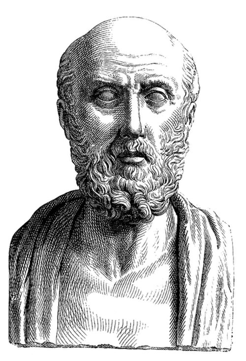
Episodes of stroke and familial stroke have been reported from the 2nd millennium BC onward in ancient Mesopotamia and Persia.[249] Hippocrates (460 to 370 BC) was first to describe the phenomenon of sudden paralysis that is often associated with ischemia. Apoplexy, from the Greek word meaning "struck down with violence", first appeared in Hippocratic writings to describe this phenomenon.[250][251] The word stroke was used as a synonym for apoplectic seizure as early as 1599,[252] and is a fairly literal translation of the Greek term. The term apoplectic stroke is an archaic, nonspecific term, for a cerebrovascular accident accompanied by haemorrhage or haemorrhagic stroke.[253] Martin Luther was described as having an apoplectic stroke that deprived him of his speech shortly before his death in 1546.[254]
In 1658, in his Apoplexia, Johann Jacob Wepfer (1620–1695) identified the cause of hemorrhagic stroke when he suggested that people who had died of apoplexy had bleeding in their brains.[52][250] Wepfer also identified the main arteries supplying the brain, the vertebral and carotid arteries, and identified the cause of a type of ischemic stroke known as a cerebral infarction when he suggested that apoplexy might be caused by a blockage to those vessels.[52] Rudolf Virchow first described the mechanism of thromboembolism as a major factor.[255]
The term cerebrovascular accident was introduced in 1927, reflecting a "growing awareness and acceptance of vascular theories and (...) recognition of the consequences of a sudden disruption in the vascular supply of the brain".[256] Its use is now discouraged by a number of neurology textbooks, reasoning that the connotation of fortuitousness carried by the word accident insufficiently highlights the modifiability of the underlying risk factors.[257][258][259] Cerebrovascular insult may be used interchangeably.[260]
The term brain attack was introduced for use to underline the acute nature of stroke according to the American Stroke Association,[260] which has used the term since 1990,[261] and is used colloquially to refer to both ischemic as well as hemorrhagic stroke.[262]
Research
As of 2017, angioplasty and stents were under preliminary clinical research to determine the possible therapeutic advantages of these procedures in comparison to therapy with statins, antithrombotics, or antihypertensive drugs.[263]
See also
References
- ^ Gaillard F. "Ischaemic stroke". radiopaedia.org. Retrieved 3 June 2018.
- ^ a b c d e f g h i j k l m n o p q r s t Donnan GA, Fisher M, Macleod M, Davis SM (May 2008). "Stroke". Lancet. 371 (9624): 1612–23. doi:10.1016/S0140-6736(08)60694-7. PMID 18468545. S2CID 208787942.(subscription required)
- ^ a b c d e f "What Are the Signs and Symptoms of a Stroke?". www.nhlbi.nih.gov. March 26, 2014. Archived from the original on 27 February 2015. Retrieved 27 February 2015.
- ^ Martin G (2009). Palliative Care Nursing: Quality Care to the End of Life, Third Edition. Springer Publishing Company. p. 290. ISBN 978-0-8261-5792-8. Archived from the original on 2017-08-03.
- ^ a b c d e f g "What Is a Stroke?". www.nhlbi.nih.gov/. March 26, 2014. Archived from the original on 18 February 2015. Retrieved 26 February 2015.
- ^ "Stroke - Causes". 24 October 2017.
- ^ a b c "Who Is at Risk for a Stroke?". www.nhlbi.nih.gov. March 26, 2014. Archived from the original on 27 February 2015. Retrieved 27 February 2015.
- ^ a b c Hu A, Niu J, Winkelmayer WC (November 2018). "Oral Anticoagulation in Patients With End-Stage Kidney Disease on Dialysis and Atrial Fibrillation". Seminars in Nephrology. 38 (6): 618–628. doi:10.1016/j.semnephrol.2018.08.006. PMC 6233322. PMID 30413255.
- ^ a b c d e "How Is a Stroke Diagnosed?". www.nhlbi.nih.gov. March 26, 2014. Archived from the original on 27 February 2015. Retrieved 27 February 2015.
- ^ a b c d Yew KS, Cheng E (July 2009). "Acute stroke diagnosis". American Family Physician. 80 (1): 33–40. PMC 2722757. PMID 19621844.
- ^ a b GBD 2015 Disease and Injury Incidence and Prevalence Collaborators (October 2016). "Global, regional, and national incidence, prevalence, and years lived with disability for 310 diseases and injuries, 1990-2015: a systematic analysis for the Global Burden of Disease Study 2015". Lancet. 388 (10053): 1545–1602. doi:10.1016/S0140-6736(16)31678-6. PMC 5055577. PMID 27733282.
- ^ a b c GBD 2015 Mortality and Causes of Death Collaborators (October 2016). "Global, regional, and national life expectancy, all-cause mortality, and cause-specific mortality for 249 causes of death, 1980-2015: a systematic analysis for the Global Burden of Disease Study 2015". Lancet. 388 (10053): 1459–1544. doi:10.1016/S0140-6736(16)31012-1. PMC 5388903. PMID 27733281.
- ^ a b c "Types of Stroke". www.nhlbi.nih.gov. March 26, 2014. Archived from the original on 19 March 2015. Retrieved 27 February 2015.
- ^ Roos KL (2012). Emergency Neurology. Springer Science & Business Media. p. 360. ISBN 978-0-387-88584-1. Archived from the original on 2017-01-08.
- ^ Wityk RJ, Llinas RH (2007). Stroke. ACP Press. p. 296. ISBN 978-1-930513-70-9. Archived from the original on 2017-01-08.
- ^ Feigin VL, Rinkel GJ, Lawes CM, Algra A, Bennett DA, van Gijn J, et al. (December 2005). "Risk factors for subarachnoid hemorrhage: an updated systematic review of epidemiological studies". Stroke. 36 (12): 2773–80. doi:10.1161/01.STR.0000190838.02954.e8. PMID 16282541.
- ^ "Stroke, cerebrovascular accident". World Health Organization. 2024. Retrieved 12 March 2024.
- ^ "The top 10 causes of death". www.who.int. Retrieved 2024-08-12.
- ^ a b c d e Feigin VL, Forouzanfar MH, Krishnamurthi R, Mensah GA, Connor M, Bennett DA, et al. (January 2014). "Global and regional burden of stroke during 1990-2010: findings from the Global Burden of Disease Study 2010". Lancet. 383 (9913): 245–54. doi:10.1016/S0140-6736(13)61953-4. PMC 4181600. PMID 24449944.
- ^ "Brain Basics: Preventing Stroke". National Institute of Neurological Disorders and Stroke. Archived from the original on 2009-10-08. Retrieved 2009-10-24.
- ^ World Health Organisation (1978). Cerebrovascular Disorders (Offset Publications). Geneva: World Health Organization. ISBN 978-92-4-170043-6. OCLC 4757533.
- ^ Kidwell CS, Warach S (December 2003). "Acute ischemic cerebrovascular syndrome: diagnostic criteria". Stroke. 34 (12): 2995–8. doi:10.1161/01.STR.0000098902.69855.A9. PMID 14605325. S2CID 16083887.
- ^ Shuaib A, Hachinski VC (September 1991). "Mechanisms and management of stroke in the elderly". CMAJ. 145 (5): 433–43. PMC 1335826. PMID 1878825.
- ^ a b Stam J (April 2005). "Thrombosis of the cerebral veins and sinuses" (PDF). The New England Journal of Medicine. 352 (17): 1791–8. doi:10.1056/NEJMra042354. PMID 15858188. S2CID 42126852.
- ^ Guercini F, Acciarresi M, Agnelli G, Paciaroni M (April 2008). "Cryptogenic stroke: time to determine aetiology". Journal of Thrombosis and Haemostasis. 6 (4): 549–54. doi:10.1111/j.1538-7836.2008.02903.x. PMID 18208534. S2CID 20211745.
- ^ Bamford J, Sandercock P, Dennis M, Burn J, Warlow C (June 1991). "Classification and natural history of clinically identifiable subtypes of cerebral infarction". Lancet. 337 (8756): 1521–6. doi:10.1016/0140-6736(91)93206-O. PMID 1675378. S2CID 21784682. Later publications distinguish between "syndrome" and "infarct", based on evidence from imaging. "Syndrome" may be replaced by "hemorrhage" if imaging demonstrates a bleed. See Internet Stroke Center. "Oxford Stroke Scale". Archived from the original on 2008-10-25. Retrieved 2008-11-14.
- ^ Bamford JM (2000). "The role of the clinical examination in the subclassification of stroke". Cerebrovascular Diseases. 10 (4): 2–4. doi:10.1159/000047582. PMID 11070389. S2CID 29493084.
- ^ Adams HP, Bendixen BH, Kappelle LJ, Biller J, Love BB, Gordon DL, et al. (January 1993). "Classification of subtype of acute ischemic stroke. Definitions for use in a multicenter clinical trial. TOAST. Trial of Org 10172 in Acute Stroke Treatment". Stroke. 24 (1): 35–41. doi:10.1161/01.STR.24.1.35. PMID 7678184.
- ^ Osterweil N (26 December 2006). "Methamphetamine May Trigger Ischemic Stroke". Medpage Today.
- ^ Anonymous (2014-07-29). "Hemorrhagic stroke". National Stroke Association. Archived from the original on 27 June 2016. Retrieved 30 June 2016.
- ^ Anonymous (6 December 2013). "Types of Stroke". www.cdc.gov. Centers of Disease Control and Prevention. Archived from the original on 27 June 2016. Retrieved 30 June 2016.
- ^ Al-Shahi Salman R, Labovitz DL, Stapf C (July 2009). "Spontaneous intracerebral haemorrhage". BMJ. 339 (jul24 1): b2586. doi:10.1136/bmj.b2586. PMID 19633038. S2CID 206891608.
- ^ a b "Articles". Cedars-Sinai. Archived from the original on 2020-05-30. Retrieved 2022-04-26.
- ^ Goldstein LB, Simel DL (May 2005). "Is this patient having a stroke?". JAMA. 293 (19): 2391–402. doi:10.1001/jama.293.19.2391. PMID 15900010. S2CID 20408776.
- ^ Harbison J, Massey A, Barnett L, Hodge D, Ford GA (June 1999). "Rapid ambulance protocol for acute stroke". Lancet. 353 (9168): 1935. doi:10.1016/S0140-6736(99)00966-6. PMID 10371574. S2CID 36692451.
- ^ Merwick Á, Werring D (May 2014). "Posterior circulation ischaemic stroke" (PDF). BMJ. 348 (may19 33): g3175. doi:10.1136/bmj.g3175. PMID 24842277.
- ^ Aroor S, Singh R, Goldstein LB (February 2017). "BE-FAST (Balance, Eyes, Face, Arm, Speech, Time): Reducing the Proportion of Strokes Missed Using the FAST Mnemonic". Stroke. 48 (2): 479–481. doi:10.1161/STROKEAHA.116.015169. PMID 28082668.
- ^ Chen X, Zhao X, Xu F, Guo M, Yang Y, Zhong L, et al. (2021). "A Systematic Review and Meta-Analysis Comparing FAST and BEFAST in Acute Stroke Patients". Front Neurol. 12: 765069. doi:10.3389/fneur.2021.765069. PMC 8837419. PMID 35153975.
- ^ Kidwell CS, Saver JL, Schubert GB, Eckstein M, Starkman S (1998). "Design and retrospective analysis of the Los Angeles Prehospital Stroke Screen (LAPSS)". Prehospital Emergency Care. 2 (4): 267–73. doi:10.1080/10903129808958878. PMID 9799012.
- ^ Kothari RU, Pancioli A, Liu T, Brott T, Broderick J (April 1999). "Cincinnati Prehospital Stroke Scale: reproducibility and validity". Annals of Emergency Medicine. 33 (4): 373–8. doi:10.1016/S0196-0644(99)70299-4. PMID 10092713.
- ^ Harbison J, Hossain O, Jenkinson D, Davis J, Louw SJ, Ford GA (January 2003). "Diagnostic accuracy of stroke referrals from primary care, emergency room physicians, and ambulance staff using the face arm speech test". Stroke. 34 (1): 71–6. doi:10.1161/01.str.0000044170.46643.5e. PMID 12511753.
- ^ a b National Institute for Health and Clinical Excellence. Clinical guideline 68: Stroke. London, 2008.
- ^ Nor AM, Davis J, Sen B, Shipsey D, Louw SJ, Dyker AG, et al. (November 2005). "The Recognition of Stroke in the Emergency Room (ROSIER) scale: development and validation of a stroke recognition instrument". The Lancet. Neurology. 4 (11): 727–34. doi:10.1016/S1474-4422(05)70201-5. PMID 16239179. S2CID 2744751.
- ^ a b c d e f O'Sullivan SB (2007). "Stroke". In O'Sullivan SB, Schmitz TJ (eds.). Physical Rehabilitation. Vol. 5. Philadelphia: F.A. Davis Company. p. 719.
- ^ Lebedeva ER, Ushenin AV, Gurary NM, Gilev DV, Olesen J (January 2022). "Diagnostic criteria for acute headache attributed to ischemic stroke and for sentinel headache before ischemic stroke". The Journal of Headache and Pain. 23 (1): 11. doi:10.1186/s10194-021-01372-x. PMC 8903596. PMID 35057731.
- ^ MedlinePlus Encyclopedia: Thrombus
- ^ "Circle of Willis". The Internet Stroke Center. Archived from the original on 2016-02-05.
- ^ Correia PN, Meyer IA, Eskandari A, Michel P (April 2016). "Beauty parlor stroke revisited: An 11-year single-center consecutive series". International Journal of Stroke. 11 (3): 356–360. doi:10.1177/1747493015620809. PMID 26763920. S2CID 36985608.
- ^ "Brain anaurysm – Introduction". NHS Choices. 2017-10-19. Archived from the original on 2016-02-08.
- ^ Fisher CM (December 1968). "The arterial lesions underlying lacunes". Acta Neuropathologica. 12 (1): 1–15. doi:10.1007/BF00685305. PMID 5708546. S2CID 6942826.
- ^ Heo J, Youk TM, Seo KD (June 2021). "Anemia Is a Risk Factor for the Development of Ischemic Stroke and Post-Stroke Mortality". Journal of Clinical Medicine. 10 (12): 2556. doi:10.3390/jcm10122556. PMC 8226740. PMID 34207841.
- ^ a b c d e f g National Institute of Neurological Disorders and Stroke (NINDS) (1999). "Stroke: Hope Through Research". National Institutes of Health. Archived from the original on 2015-10-04.
- ^ Shah AS, Lee KK, McAllister DA, Hunter A, Nair H, Whiteley W, et al. (March 2015). "Short term exposure to air pollution and stroke: systematic review and meta-analysis". BMJ. 350 (mar23 11): h1295. doi:10.1136/bmj.h1295. PMC 4373601. PMID 25810496.
- ^ a b Kumar V (2009). Robbins and Cotran Pathologic Basis of Disease, Professional Edition (8th ed.). Philadelphia: Elsevier. ISBN 978-1-4377-0792-2.
- ^ Ay H, Furie KL, Singhal A, Smith WS, Sorensen AG, Koroshetz WJ (November 2005). "An evidence-based causative classification system for acute ischemic stroke". Annals of Neurology. 58 (5): 688–97. doi:10.1002/ana.20617. PMID 16240340. S2CID 28798146.
- ^ Hackam DG (May 2016). "Prognosis of Asymptomatic Carotid Artery Occlusion: Systematic Review and Meta-Analysis". Stroke. 47 (5): 1253–7. doi:10.1161/strokeaha.116.012760. PMID 27073237. S2CID 3669224.
- ^ Hart RG, Catanese L, Perera KS, Ntaios G, Connolly SJ (April 2017). "Embolic Stroke of Undetermined Source: A Systematic Review and Clinical Update". Stroke. 48 (4): 867–872. doi:10.1161/STROKEAHA.116.016414. PMID 28265016. S2CID 3679562.
- ^ Strandgaard S (October 1996). "Hypertension and stroke". Journal of Hypertension Supplement. 14 (3): S23-7. doi:10.1097/00004872-199610003-00005. PMID 9120662. S2CID 11817729.
- ^ Harrigan MR, Deveikis JP (2012). Handbook of Cerebrovascular Disease and Neurointerventional Technique. Springer Science & Business Media. p. 692. ISBN 978-1-61779-945-7. Archived from the original on 2017-01-09.
- ^ Navi BB, Iadecola C (May 2018). "Ischemic stroke in cancer patients: A review of an underappreciated pathology". Annals of Neurology. 83 (5): 873–883. doi:10.1002/ana.25227. PMC 6021225. PMID 29633334.
- ^ Miwa K, Hoshi T, Hougaku H, Tanaka M, Furukado S, Abe Y, et al. (2010). "Silent cerebral infarction is associated with incident stroke and TIA independent of carotid intima-media thickness". Internal Medicine. 49 (9): 817–22. doi:10.2169/internalmedicine.49.3211. PMID 20453400.
- ^ a b Herderscheê D, Hijdra A, Algra A, Koudstaal PJ, Kappelle LJ, van Gijn J (September 1992). "Silent stroke in patients with transient ischemic attack or minor ischemic stroke. The Dutch TIA Trial Study Group". Stroke. 23 (9): 1220–4. doi:10.1161/01.STR.23.9.1220. PMID 1519274.
- ^ Leary MC, Saver JL (2003). "Annual incidence of first silent stroke in the United States: a preliminary estimate". Cerebrovascular Diseases. 16 (3): 280–5. doi:10.1159/000071128. PMID 12865617. S2CID 33095581.
- ^ Vermeer SE, Koudstaal PJ, Oudkerk M, Hofman A, Breteler MM (January 2002). "Prevalence and risk factors of silent brain infarcts in the population-based Rotterdam Scan Study". Stroke. 33 (1): 21–5. doi:10.1161/hs0102.101629. PMID 11779883.
- ^ Deb P, Sharma S, Hassan KM (June 2010). "Pathophysiologic mechanisms of acute ischemic stroke: An overview with emphasis on therapeutic significance beyond thrombolysis". Pathophysiology. 17 (3): 197–218. doi:10.1016/j.pathophys.2009.12.001. PMID 20074922.
- ^ Snell RS (2006). Clinical neuroanatomy, 6. ed. Lippincott Williams & Wilkins, Philadelphia. pp. 478–85. ISBN 978-963-226-293-2.
- ^ Brunner and Suddarth's Textbook on Medical-Surgical Nursing, 11th Edition
- ^ Lo EH (2008). "A new penumbra: transitioning from injury into repair after stroke". Nature Medicine. 14 (5): 497–500. doi:10.1038/nm1735. PMID 18463660. S2CID 205385488.
- ^ Kristián T, Siesjö BK (1996). "Calcium-related damage in ischemia". Life Sciences. 59 (5–6): 357–67. doi:10.1016/0024-3205(96)00314-1. PMID 8761323.
- ^ a b "Ischemic Stroke - Neurologic Disorders". MSD Manual Professional Edition. Retrieved 2023-02-14.
- ^ Iwasawa E, Ichijo M, Ishibashi S, Yokota T (March 2016). "Acute development of collateral circulation and therapeutic prospects in ischemic stroke". Neural Regeneration Research. 11 (3): 368–371. doi:10.4103/1673-5374.179033. PMC 4828985. PMID 27127459.
- ^ Liu L, Ding J, Leng X, Pu Y, Huang LA, Xu A, et al. (September 2018). "Guidelines for evaluation and management of cerebral collateral circulation in ischaemic stroke 2017". Stroke and Vascular Neurology. 3 (3): 117–130. doi:10.1136/svn-2017-000135. PMC 6169613. PMID 30294467.
- ^ a b Smith WS, English JD, Johnston SC (2012). "Chapter 370: Cerebrovascular Diseases". In Longo DL, Fauci AS, Kasper DL, Hauser SL, Jameson JL, Loscalzo J (eds.). Harrison's principles of internal medicine (18th ed.). New York: McGraw-Hill. pp. 3270–3299. ISBN 978-0-07-174889-6.
- ^ a b Wang J (December 2010). "Preclinical and clinical research on inflammation after intracerebral hemorrhage". Progress in Neurobiology. 92 (4): 463–77. doi:10.1016/j.pneurobio.2010.08.001. PMC 2991407. PMID 20713126.
- ^ Hill MD (November 2005). "Diagnostic biomarkers for stroke: a stroke neurologist's perspective". Clinical Chemistry. 51 (11): 2001–2. doi:10.1373/clinchem.2005.056382. PMID 16244286.
- ^ Chalela JA, Kidwell CS, Nentwich LM, Luby M, Butman JA, Demchuk AM, et al. (January 2007). "Magnetic resonance imaging and computed tomography in emergency assessment of patients with suspected acute stroke: a prospective comparison". Lancet. 369 (9558): 293–8. doi:10.1016/S0140-6736(07)60151-2. PMC 1859855. PMID 17258669.
- ^ Kidwell CS, Chalela JA, Saver JL, Starkman S, Hill MD, Demchuk AM, et al. (October 2004). "Comparison of MRI and CT for detection of acute intracerebral hemorrhage". JAMA. 292 (15): 1823–30. doi:10.1001/jama.292.15.1823. PMID 15494579.
- ^ Nouh A, Remke J, Ruland S (2014). "Ischemic posterior circulation stroke: a review of anatomy, clinical presentations, diagnosis, and current management". Frontiers in Neurology. 5: 30. doi:10.3389/fneur.2014.00030. PMC 3985033. PMID 24778625.
- ^ Tong E, Patrie J, Tong S, Evans A, Michel P, Eskandari A, et al. (November 2017). "Time-resolved CT assessment of collaterals as imaging biomarkers to predict clinical outcomes in acute ischemic stroke". Neuroradiology. 59 (11): 1101–1109. doi:10.1007/s00234-017-1914-z. PMID 28864854. S2CID 19070631.
- ^ Stoll M, Hamann GF (August 2002). "[Cerebrovascular reserve capacity]". Der Nervenarzt (in German). 73 (8): 711–8. doi:10.1007/s00115-002-1313-4. PMID 12242957. S2CID 34870086.
- ^ Sebök M, Niftrik BV, Lohaus N, Esposito G, Amki ME, Winklhofer S, et al. (November 2021). "Leptomeningeal collateral activation indicates severely impaired cerebrovascular reserve capacity in patients with symptomatic unilateral carotid artery occlusion". Journal of Cerebral Blood Flow and Metabolism. 41 (11): 3039–3051. doi:10.1177/0271678X211024373. PMC 8545056. PMID 34112002.
- ^ a b Wilson D, Adams ME, Robertson F, Murphy M, Werring DJ (May 2015). "Investigating intracerebral haemorrhage". BMJ. 350 (may20 10): h2484. doi:10.1136/bmj.h2484. PMID 25994363. S2CID 26908106.
- ^ a b c d e Bakradze E, Liberman AL (February 2018). "Diagnostic Error in Stroke-Reasons and Proposed Solutions". Current Atherosclerosis Reports. 20 (2): 11. doi:10.1007/s11883-018-0712-3. PMID 29441421. S2CID 3335617.
- ^ Dupre CM, Libman R, Dupre SI, Katz JM, Rybinnik I, Kwiatkowski T (February 2014) [Available online 15 August 2013]. "Stroke chameleons". Journal of Stroke and Cerebrovascular Diseases. 23 (2): 374–8. doi:10.1016/j.jstrokecerebrovasdis.2013.07.015. PMID 23954604.
- ^ a b Straus SE, Majumdar SR, McAlister FA (September 2002). "New evidence for stroke prevention: scientific review". JAMA. 288 (11): 1388–95. doi:10.1001/jama.288.11.1388. PMID 12234233.
- ^ Goldstein LB, Adams R, Alberts MJ, Appel LJ, Brass LM, Bushnell CD, et al. (June 2006). "Primary prevention of ischemic stroke: a guideline from the American Heart Association/American Stroke Association Stroke Council: cosponsored by the Atherosclerotic Peripheral Vascular Disease Interdisciplinary Working Group; Cardiovascular Nursing Council; Clinical Cardiology Council; Nutrition, Physical Activity, and Metabolism Council; and the Quality of Care and Outcomes Research Interdisciplinary Working Group: the American Academy of Neurology affirms the value of this guideline". Stroke. 37 (6): 1583–633. doi:10.1161/01.STR.0000223048.70103.F1. PMID 16675728.
- ^ Center for Drug Evaluation and Research. "Information for Consumers (Drugs) - Use of Aspirin for Primary Prevention of Heart Attack and Stroke". www.fda.gov. Archived from the original on 2015-11-17. Retrieved 2015-11-16.
- ^ a b c "Stroke prevention - Antiplatelets and anticoagulants: NPS - Better choices, Better health". 2012-04-07. Archived from the original on 2012-04-07. Retrieved 2024-01-29.
{{cite web}}: CS1 maint: bot: original URL status unknown (link) - ^ a b Antithrombotic Trialists' Collaboration (January 2002). "Collaborative meta-analysis of randomised trials of antiplatelet therapy for prevention of death, myocardial infarction, and stroke in high risk patients". BMJ. 324 (7329): 71–86. doi:10.1136/bmj.324.7329.71. PMC 64503. PMID 11786451.
- ^ Jonas DE, Feltner C, Amick HR, Sheridan S, Zheng ZJ, Watford DJ, et al. (September 2014). "Screening for asymptomatic carotid artery stenosis: a systematic review and meta-analysis for the U.S. Preventive Services Task Force". Annals of Internal Medicine. 161 (5): 336–46. doi:10.7326/M14-0530. PMID 25004169. S2CID 8741746.
- ^ Medical Research Council Working Party (July 1985). "MRC trial of treatment of mild hypertension: principal results. Medical Research Council Working Party". British Medical Journal. 291 (6488): 97–104. doi:10.1136/bmj.291.6488.97. PMC 1416260. PMID 2861880.
- ^ Thomson R (August 2009). "Evidence based implementation of complex interventions". BMJ. 339: b3124. doi:10.1136/bmj.b3124. PMID 19675081. S2CID 692596.
- ^ Hankey GJ (August 1999). "Smoking and risk of stroke". Journal of Cardiovascular Risk. 6 (4): 207–11. doi:10.1177/204748739900600403. PMID 10501270. S2CID 43819614.
- ^ Wannamethee SG, Shaper AG, Whincup PH, Walker M (July 1995). "Smoking cessation and the risk of stroke in middle-aged men". JAMA. 274 (2): 155–60. doi:10.1001/jama.274.2.155. PMID 7596004.
- ^ Reynolds K, Lewis B, Nolen JD, Kinney GL, Sathya B, He J, et al. (February 2003). "Alcohol consumption and risk of stroke: a meta-analysis". JAMA. 289 (5): 579–88. doi:10.1001/jama.289.5.579. PMID 12578491. S2CID 28076015.
- ^ Sloan MA, Kittner SJ, Rigamonti D, Price TR (September 1991). "Occurrence of stroke associated with use/abuse of drugs". Neurology. 41 (9): 1358–64. doi:10.1212/WNL.41.9.1358. PMID 1891081. S2CID 26670239.
- ^ Larsson SC, Virtamo J, Wolk A (August 2011). "Red meat consumption and risk of stroke in Swedish men". The American Journal of Clinical Nutrition. 94 (2): 417–21. doi:10.3945/ajcn.111.015115. PMID 21653800.
- ^ "Stroke Risk Factors". American Heart Association. 2007. Retrieved January 22, 2007.
- ^ Hackshaw A, Morris JK, Boniface S, Tang JL, Milenković D (January 2018). "Low cigarette consumption and risk of coronary heart disease and stroke: meta-analysis of 141 cohort studies in 55 study reports". BMJ. 360: j5855. doi:10.1136/bmj.j5855. PMC 5781309. PMID 29367388.
- ^ Gorelick PB (1987). "Alcohol and stroke". Stroke. 18 (1): 268–71. doi:10.1161/01.STR.18.1.268. PMID 3810763.
- ^ Westover AN, McBride S, Haley RW (April 2007). "Stroke in young adults who abuse amphetamines or cocaine: a population-based study of hospitalized patients". Archives of General Psychiatry. 64 (4): 495–502. doi:10.1001/archpsyc.64.4.495. PMID 17404126.
- ^ Schürks M, Rist PM, Bigal ME, Buring JE, Lipton RB, Kurth T (October 2009). "Migraine and cardiovascular disease: systematic review and meta-analysis". BMJ. 339 (oct27 1): b3914. doi:10.1136/bmj.b3914. PMC 2768778. PMID 19861375.
- ^ Kurth T, Chabriat H, Bousser MG (January 2012). "Migraine and stroke: a complex association with clinical implications". The Lancet. Neurology. 11 (1): 92–100. doi:10.1016/S1474-4422(11)70266-6. PMID 22172624. S2CID 31939284.
- ^ Ciaccio EJ, Lewis SK, Biviano AB, Iyer V, Garan H, Green PH (August 2017). "Cardiovascular involvement in celiac disease". World Journal of Cardiology (Review). 9 (8): 652–666. doi:10.4330/wjc.v9.i8.652. PMC 5583538. PMID 28932354.
- ^ Pega F, Náfrádi B, Momen NC, Ujita Y, Streicher KN, Prüss-Üstün AM, et al. (September 2021). "Global, regional, and national burdens of ischemic heart disease and stroke attributable to exposure to long working hours for 194 countries, 2000-2016: A systematic analysis from the WHO/ILO Joint Estimates of the Work-related Burden of Disease and Injury". Environment International. 154: 106595. Bibcode:2021EnInt.15406595P. doi:10.1016/j.envint.2021.106595. PMC 8204267. PMID 34011457.
- ^ Kyu HH, Bachman VF, Alexander LT, Mumford JE, Afshin A, Estep K, et al. (August 2016). "Physical activity and risk of breast cancer, colon cancer, diabetes, ischemic heart disease, and ischemic stroke events: systematic review and dose-response meta-analysis for the Global Burden of Disease Study 2013". BMJ. 354: i3857. doi:10.1136/bmj.i3857. PMC 4979358. PMID 27510511.
- ^ Ezekowitz JA, Straus SE, Majumdar SR, McAlister FA (December 2003). "Stroke: strategies for primary prevention". American Family Physician. 68 (12): 2379–86. PMID 14705756.
- ^ a b c d Ederle J, Brown MM (October 2006). "The evidence for medicine versus surgery for carotid stenosis". European Journal of Radiology. 60 (1): 3–7. doi:10.1016/j.ejrad.2006.05.021. PMID 16920313.
- ^ Whisnant JP (February 1996). "Effectiveness versus efficacy of treatment of hypertension for stroke prevention". Neurology. 46 (2): 301–7. doi:10.1212/WNL.46.2.301. PMID 8614485. S2CID 28985425.
- ^ Law MR, Morris JK, Wald NJ (May 2009). "Use of blood pressure lowering drugs in the prevention of cardiovascular disease: meta-analysis of 147 randomised trials in the context of expectations from prospective epidemiological studies". BMJ. 338: b1665. doi:10.1136/bmj.b1665. PMC 2684577. PMID 19454737.
- ^ Psaty BM, Lumley T, Furberg CD, Schellenbaum G, Pahor M, Alderman MH, et al. (May 2003). "Health outcomes associated with various antihypertensive therapies used as first-line agents: a network meta-analysis". JAMA. 289 (19): 2534–44. doi:10.1001/jama.289.19.2534. PMID 12759325. S2CID 123289.
- ^ a b "Cholesterol, diastolic blood pressure, and stroke: 13,000 strokes in 450,000 people in 45 prospective cohorts. Prospective studies collaboration". Lancet. 346 (8991–8992): 1647–53. 1995. doi:10.1016/S0140-6736(95)92836-7. PMID 8551820. S2CID 12043767.
- ^ Gueyffier F, Boissel JP, Boutitie F, Pocock S, Coope J, Cutler J, et al. (December 1997). "Effect of antihypertensive treatment in patients having already suffered from stroke. Gathering the evidence. The INDANA (INdividual Data ANalysis of Antihypertensive intervention trials) Project Collaborators". Stroke. 28 (12): 2557–62. doi:10.1161/01.STR.28.12.2557. PMID 9412649.
- ^ Gueyffier F, Bulpitt C, Boissel JP, Schron E, Ekbom T, Fagard R, et al. (March 1999). "Antihypertensive drugs in very old people: a subgroup meta-analysis of randomised controlled trials. INDANA Group". Lancet. 353 (9155): 793–6. doi:10.1016/S0140-6736(98)08127-6. PMID 10459960. S2CID 43858004.
- ^ Staessen JA, Gasowski J, Wang JG, Thijs L, Den Hond E, Boissel JP, et al. (March 2000). "Risks of untreated and treated isolated systolic hypertension in the elderly: meta-analysis of outcome trials". Lancet. 355 (9207): 865–72. doi:10.1016/S0140-6736(99)07330-4. PMID 10752701. S2CID 31403774.
- ^ Beckett NS, Peters R, Fletcher AE, Staessen JA, Liu L, Dumitrascu D, et al. (May 2008). "Treatment of hypertension in patients 80 years of age or older" (PDF). The New England Journal of Medicine. 358 (18): 1887–98. doi:10.1056/NEJMoa0801369. PMID 18378519. Archived (PDF) from the original on November 8, 2023.
- ^ Neal B, MacMahon S, Chapman N (December 2000). "Effects of ACE inhibitors, calcium antagonists, and other blood-pressure-lowering drugs: results of prospectively designed overviews of randomised trials. Blood Pressure Lowering Treatment Trialists' Collaboration". Lancet. 356 (9246): 1955–64. doi:10.1016/S0140-6736(00)03307-9. PMID 11130523. S2CID 46148907.
- ^ The Allhat Officers and Coordinators for the Allhat Collaborative Research Group (December 2002). "Major outcomes in high-risk hypertensive patients randomized to angiotensin-converting enzyme inhibitor or calcium channel blocker vs diuretic: The Antihypertensive and Lipid-Lowering Treatment to Prevent Heart Attack Trial (ALLHAT)". JAMA. 288 (23): 2981–97. doi:10.1001/jama.288.23.2981. PMID 12479763.
- ^ De Lima LG, Saconato H, Atallah AN, da Silva EM (October 2014). "Beta-blockers for preventing stroke recurrence". The Cochrane Database of Systematic Reviews. 2014 (10): CD007890. doi:10.1002/14651858.CD007890.pub3. PMC 10590202. PMID 25317988.
- ^ Iso H, Jacobs DR, Wentworth D, Neaton JD, Cohen JD (April 1989). "Serum cholesterol levels and six-year mortality from stroke in 350,977 men screened for the multiple risk factor intervention trial". The New England Journal of Medicine. 320 (14): 904–10. doi:10.1056/NEJM198904063201405. PMID 2619783.
- ^ a b O'Regan C, Wu P, Arora P, Perri D, Mills EJ (January 2008). "Statin therapy in stroke prevention: a meta-analysis involving 121,000 patients". The American Journal of Medicine. 121 (1): 24–33. doi:10.1016/j.amjmed.2007.06.033. PMID 18187070.
- ^ Hebert PR, Gaziano JM, Hennekens CH (January 1995). "An overview of trials of cholesterol lowering and risk of stroke". Archives of Internal Medicine. 155 (1): 50–5. doi:10.1001/archinte.155.1.50. PMID 7802520.
- ^ "Intensive blood-glucose control with sulphonylureas or insulin compared with conventional treatment and risk of complications in patients with type 2 diabetes (UKPDS 33). UK Prospective Diabetes Study (UKPDS) Group". Lancet. 352 (9131): 837–53. September 1998. doi:10.1016/S0140-6736(98)07019-6. PMID 9742976. S2CID 7019505.
- ^ Dormandy JA, Charbonnel B, Eckland DJ, Erdmann E, Massi-Benedetti M, Moules IK, et al. (October 2005). "Secondary prevention of macrovascular events in patients with type 2 diabetes in the PROactive Study (PROspective pioglitAzone Clinical Trial In macroVascular Events): a randomised controlled trial". Lancet. 366 (9493): 1279–89. doi:10.1016/S0140-6736(05)67528-9. hdl:2268/6536. PMID 16214598. S2CID 11825315.
- ^ Johnson ES, Lanes SF, Wentworth CE, Satterfield MH, Abebe BL, Dicker LW (June 1999). "A metaregression analysis of the dose-response effect of aspirin on stroke". Archives of Internal Medicine. 159 (11): 1248–53. doi:10.1001/archinte.159.11.1248. PMID 10371234.
- ^ a b Sudlow CL, Mason G, Maurice JB, Wedderburn CJ, Hankey GJ (October 2009). "Thienopyridine derivatives versus aspirin for preventing stroke and other serious vascular events in high vascular risk patients". The Cochrane Database of Systematic Reviews. 2009 (4): CD001246. doi:10.1002/14651858.CD001246.pub2. PMC 7055203. PMID 19821273. S2CID 205162946.
- ^ Hao Q, Tampi M, O'Donnell M, Foroutan F, Siemieniuk RA, Guyatt G (December 2018). "Clopidogrel plus aspirin versus aspirin alone for acute minor ischaemic stroke or high risk transient ischaemic attack: systematic review and meta-analysis". BMJ. 363: k5108. doi:10.1136/bmj.k5108. PMC 6298178. PMID 30563866.
- ^ Halkes PH, van Gijn J, Kappelle LJ, Koudstaal PJ, Algra A (May 2006). "Aspirin plus dipyridamole versus aspirin alone after cerebral ischaemia of arterial origin (ESPRIT): randomised controlled trial". Lancet. 367 (9523): 1665–73. doi:10.1016/S0140-6736(06)68734-5. PMID 16714187. S2CID 10691264.
- ^ Wolf PA, Abbott RD, Kannel WB (September 1987). "Atrial fibrillation: a major contributor to stroke in the elderly. The Framingham Study". Archives of Internal Medicine. 147 (9): 1561–4. doi:10.1001/archinte.147.9.1561. PMID 3632164.
- ^ Fuster V, Rydén LE, Cannom DS, Crijns HJ, Curtis AB, Ellenbogen KA, et al. (August 2006). "ACC/AHA/ESC 2006 Guidelines for the Management of Patients with Atrial Fibrillation: a report of the American College of Cardiology/American Heart Association Task Force on Practice Guidelines and the European Society of Cardiology Committee for Practice Guidelines (Writing Committee to Revise the 2001 Guidelines for the Management of Patients With Atrial Fibrillation): developed in collaboration with the European Heart Rhythm Association and the Heart Rhythm Society". Circulation. 114 (7): e257-354. doi:10.1161/CIRCULATIONAHA.106.177292. PMID 16908781.
- ^ a b Sanders GD, Lowenstern A, Borre E, Chatterjee R, Goode A, Sharan L, et al. (2018-10-30). Stroke Prevention in Patients With Atrial Fibrillation: A Systematic Review Update (Report). Agency for Healthcare Research and Quality (AHRQ). doi:10.23970/ahrqepccer214 (inactive 2024-03-18). Archived from the original on 2019-03-29. Retrieved 2023-08-20.
{{cite report}}: CS1 maint: DOI inactive as of March 2024 (link) - ^ Halkes PH, van Gijn J, Kappelle LJ, Koudstaal PJ, Algra A (February 2007). "Medium intensity oral anticoagulants versus aspirin after cerebral ischaemia of arterial origin (ESPRIT): a randomised controlled trial". The Lancet. Neurology. 6 (2): 115–24. doi:10.1016/S1474-4422(06)70685-8. PMID 17239798. S2CID 21982861.
- ^ Hart RG, Halperin JL, McBride R, Benavente O, Man-Son-Hing M, Kronmal RA (March 2000). "Aspirin for the primary prevention of stroke and other major vascular events: meta-analysis and hypotheses". Archives of Neurology. 57 (3): 326–32. doi:10.1001/archneur.57.3.326. PMID 10714657.
- ^ Bartolucci AA, Howard G (September 2006). "Meta-analysis of data from the six primary prevention trials of cardiovascular events using aspirin". The American Journal of Cardiology. 98 (6): 746–50. doi:10.1016/j.amjcard.2006.04.012. PMID 16950176.
- ^ Berger JS, Roncaglioni MC, Avanzini F, Pangrazzi I, Tognoni G, Brown DL (January 2006). "Aspirin for the primary prevention of cardiovascular events in women and men: a sex-specific meta-analysis of randomized controlled trials". JAMA. 295 (3): 306–13. doi:10.1001/jama.295.3.306. PMID 16418466. S2CID 11952921.
- ^ Yerman T, Gan WQ, Sin DD (October 2007). "The influence of gender on the effects of aspirin in preventing myocardial infarction". BMC Medicine. 5: 29. doi:10.1186/1741-7015-5-29. PMC 2131749. PMID 17949479.
- ^ Rothwell PM, Eliasziw M, Gutnikov SA, Fox AJ, Taylor DW, Mayberg MR, et al. (January 2003). "Analysis of pooled data from the randomised controlled trials of endarterectomy for symptomatic carotid stenosis". Lancet. 361 (9352): 107–16. doi:10.1016/S0140-6736(03)12228-3. PMID 12531577. S2CID 2484664.
- ^ Ringleb PA, Chatellier G, Hacke W, Favre JP, Bartoli JM, Eckstein HH, et al. (February 2008). "Safety of endovascular treatment of carotid artery stenosis compared with surgical treatment: a meta-analysis". Journal of Vascular Surgery. 47 (2): 350–5. doi:10.1016/j.jvs.2007.10.035. PMID 18241759.
- ^ Müller MD, Lyrer P, Brown MM, Bonati LH (February 2020). "Carotid artery stenting versus endarterectomy for treatment of carotid artery stenosis". The Cochrane Database of Systematic Reviews. 2020 (2): CD000515. doi:10.1002/14651858.CD000515.pub5. PMC 7041119. PMID 32096559.
- ^ Rothwell PM, Eliasziw M, Gutnikov SA, Warlow CP, Barnett HJ (March 2004). "Endarterectomy for symptomatic carotid stenosis in relation to clinical subgroups and timing of surgery". Lancet. 363 (9413): 915–24. doi:10.1016/S0140-6736(04)15785-1. PMID 15043958. S2CID 3916408.
- ^ Fairhead JF, Mehta Z, Rothwell PM (August 2005). "Population-based study of delays in carotid imaging and surgery and the risk of recurrent stroke". Neurology. 65 (3): 371–5. doi:10.1212/01.wnl.0000170368.82460.b4. PMID 16087900. S2CID 24829283.
- ^ U.S. Preventive Services Task Force (December 2007). "Screening for carotid artery stenosis: U.S. Preventive Services Task Force recommendation statement". Annals of Internal Medicine. 147 (12): 854–9. doi:10.7326/0003-4819-147-12-200712180-00005. PMID 18087056.
- ^ Halliday A, Mansfield A, Marro J, Peto C, Peto R, Potter J, et al. (May 2004). "Prevention of disabling and fatal strokes by successful carotid endarterectomy in patients without recent neurological symptoms: randomised controlled trial". Lancet. 363 (9420): 1491–502. doi:10.1016/S0140-6736(04)16146-1. PMID 15135594. S2CID 22814764.
- ^ Chambers BR, Donnan GA (October 2005). "Carotid endarterectomy for asymptomatic carotid stenosis". The Cochrane Database of Systematic Reviews. 2008 (4): CD001923. doi:10.1002/14651858.CD001923.pub2. PMC 6669257. PMID 16235289.
- ^ Spence JD (September 2006). "Nutrition and stroke prevention". Stroke. 37 (9): 2430–5. CiteSeerX 10.1.1.326.6681. doi:10.1161/01.STR.0000236633.40160.ee. PMID 16873712.
- ^ Zhou YH, Tang JY, Wu MJ, Lu J, Wei X, Qin YY, et al. (2011). "Effect of folic acid supplementation on cardiovascular outcomes: a systematic review and meta-analysis". PLOS ONE. 6 (9): e25142. Bibcode:2011PLoSO...625142Z. doi:10.1371/journal.pone.0025142. PMC 3182189. PMID 21980387.
- ^ Clarke R, Halsey J, Lewington S, Lonn E, Armitage J, Manson JE, et al. (October 2010). "Effects of lowering homocysteine levels with B vitamins on cardiovascular disease, cancer, and cause-specific mortality: Meta-analysis of 8 randomized trials involving 37 485 individuals". Archives of Internal Medicine. 170 (18): 1622–31. doi:10.1001/archinternmed.2010.348. PMID 20937919. S2CID 204988253.
- ^ Bushnell C, McCullough LD, Awad IA, Chireau MV, Fedder WN, Furie KL, et al. (May 2014). "Guidelines for the prevention of stroke in women: a statement for healthcare professionals from the American Heart Association/American Stroke Association". Stroke. 45 (5): 1545–88. doi:10.1161/01.str.0000442009.06663.48. PMC 10152977. PMID 24503673.
- ^ a b Kernan WN, Ovbiagele B, Black HR, Bravata DM, Chimowitz MI, Ezekowitz MD, et al. (July 2014). "Guidelines for the prevention of stroke in patients with stroke and transient ischemic attack: a guideline for healthcare professionals from the American Heart Association/American Stroke Association". Stroke. 45 (7): 2160–236. doi:10.1161/STR.0000000000000024. PMID 24788967.
- ^ Hart RG, Pearce LA, Aguilar MI (June 2007). "Meta-analysis: antithrombotic therapy to prevent stroke in patients who have nonvalvular atrial fibrillation". Annals of Internal Medicine. 146 (12): 857–67. doi:10.7326/0003-4819-146-12-200706190-00007. PMID 17577005. S2CID 25505238.
- ^ Paciaroni M, Agnelli G, Micheli S, Caso V (February 2007). "Efficacy and safety of anticoagulant treatment in acute cardioembolic stroke: a meta-analysis of randomized controlled trials". Stroke. 38 (2): 423–30. doi:10.1161/01.STR.0000254600.92975.1f. PMID 17204681.
- ^ Johnston SC (1 July 2007). "Review: anticoagulants increase intracerebral bleeding and do not reduce death or disability in acute cardioembolic stroke". ACP Journal Club. 147 (1): 17. doi:10.7326/ACPJC-2007-147-1-017. PMID 17608382.
- ^ Armstrong MJ, Gronseth G, Anderson DC, Biller J, Cucchiara B, Dafer R, et al. (May 2013). "Summary of evidence-based guideline: periprocedural management of antithrombotic medications in patients with ischemic cerebrovascular disease: report of the Guideline Development Subcommittee of the American Academy of Neurology". Neurology. 80 (22): 2065–9. doi:10.1212/WNL.0b013e318294b32d. PMC 3716407. PMID 23713086.
- ^ a b "Stroke Treatment". www.nhlbi.nih.gov. 26 May 2023. Archived from the original on 29 February 2024. Retrieved 18 March 2024.
- ^ Rothwell PM, Algra A, Chen Z, Diener HC, Norrving B, Mehta Z (July 2016). "Effects of aspirin on risk and severity of early recurrent stroke after transient ischaemic attack and ischaemic stroke: time-course analysis of randomised trials". Lancet. 388 (10042): 365–375. doi:10.1016/S0140-6736(16)30468-8. PMC 5321490. PMID 27209146.
- ^ Gomez CR (1993). "Editorial: Time is brain!". Journal of Stroke and Cerebrovascular Diseases. 3 (1): 1–2. doi:10.1016/S1052-3057(10)80125-9. PMID 26487071.
- ^ Saver JL (January 2006). "Time is brain--quantified". Stroke. 37 (1): 263–6. doi:10.1161/01.STR.0000196957.55928.ab. PMID 16339467.
- ^ Bellolio MF, Gilmore RM, Ganti L (January 2014). "Insulin for glycaemic control in acute ischaemic stroke". The Cochrane Database of Systematic Reviews. 1 (1): CD005346. doi:10.1002/14651858.CD005346.pub4. PMC 10770823. PMID 24453023.
- ^ Bath PM, Krishnan K (October 2014). "Interventions for deliberately altering blood pressure in acute stroke". The Cochrane Database of Systematic Reviews. 2014 (10): CD000039. doi:10.1002/14651858.CD000039.pub3. PMC 7052738. PMID 25353321.
- ^ Lee M, Ovbiagele B, Hong KS, Wu YL, Lee JE, Rao NM, et al. (July 2015). "Effect of Blood Pressure Lowering in Early Ischemic Stroke: Meta-Analysis". Stroke. 46 (7): 1883–9. doi:10.1161/STROKEAHA.115.009552. PMID 26022636. S2CID 16136213.
- ^ a b Ziganshina LE, Abakumova T, Nurkhametova D, Ivanchenko K, et al. (Cochrane Stroke Group) (October 2023). "Cerebrolysin for acute ischaemic stroke". The Cochrane Database of Systematic Reviews. 2023 (10): CD007026. doi:10.1002/14651858.CD007026.pub7. PMC 10565895. PMID 37818733.
- ^ a b c Wardlaw JM, Murray V, Berge E, del Zoppo GJ (July 2014). "Thrombolysis for acute ischaemic stroke". The Cochrane Database of Systematic Reviews. 7 (7): CD000213. doi:10.1002/14651858.CD000213.pub3. PMC 4153726. PMID 25072528.
- ^ a b Emberson J, Lees KR, Lyden P, Blackwell L, Albers G, Bluhmki E, et al. (November 2014). "Effect of treatment delay, age, and stroke severity on the effects of intravenous thrombolysis with alteplase for acute ischaemic stroke: a meta-analysis of individual patient data from randomised trials". Lancet. 384 (9958): 1929–35. doi:10.1016/S0140-6736(14)60584-5. PMC 4441266. PMID 25106063.
- ^ a b "Thrombolytics for acute stroke". Dynamed. Sep 15, 2014.
at 3–4.5 hours after stroke onset t-PA increases risk of symptomatic intracranial hemorrhage but effect on functional outcomes is inconsistent
[verification needed] - ^ Alper BS, Malone-Moses M, McLellan JS, Prasad K, Manheimer E (March 2015). "Thrombolysis in acute ischaemic stroke: time for a rethink?". BMJ. 350 (h1075): h1075. doi:10.1136/bmj.h1075. PMID 25786912. S2CID 38909467.
- ^ "Canadian Association of Emergency Physicians Position Statement on Acute Ischemic Stroke" (PDF). caep.ca. March 2015. Archived from the original (PDF) on 2015-09-18. Retrieved 7 April 2015.
- ^ "10 points to remember". American College of Cardiology. 2018. Retrieved March 27, 2020.
- ^ Wardlaw JM, Murray V, Berge E, del Zoppo G, Sandercock P, Lindley RL, et al. (June 2012). "Recombinant tissue plasminogen activator for acute ischaemic stroke: an updated systematic review and meta-analysis". Lancet. 379 (9834): 2364–72. doi:10.1016/S0140-6736(12)60738-7. PMC 3386494. PMID 22632907.
- ^ Whiteley WN, Slot KB, Fernandes P, Sandercock P, Wardlaw J (November 2012). "Risk factors for intracranial hemorrhage in acute ischemic stroke patients treated with recombinant tissue plasminogen activator: a systematic review and meta-analysis of 55 studies". Stroke. 43 (11): 2904–9. doi:10.1161/STROKEAHA.112.665331. PMID 22996959.
- ^ Campbell BC, Ma H, Ringleb PA, Parsons MW, Churilov L, Bendszus M, et al. (July 2019). "Extending thrombolysis to 4·5-9 h and wake-up stroke using perfusion imaging: a systematic review and meta-analysis of individual patient data". Lancet. 394 (10193): 139–147. doi:10.1016/S0140-6736(19)31053-0. hdl:10138/312914. PMID 31128925. S2CID 205990717.
- ^ The National Institute Of Neurological Disorders And Stroke Rt-Pa Stroke Study Group (December 1995). "Tissue plasminogen activator for acute ischemic stroke". The New England Journal of Medicine. 333 (24): 1581–7. doi:10.1056/NEJM199512143332401. PMID 7477192.
- ^ Wardlaw JM, Murray V, Berge E, del Zoppo GJ (July 2014). "Thrombolysis for acute ischaemic stroke". The Cochrane Database of Systematic Reviews. 7 (7): CD000213. doi:10.1002/14651858.CD000213.pub3. PMC 4153726. PMID 25072528.
- ^ Dubinsky R, Lai SM (June 2006). "Mortality of stroke patients treated with thrombolysis: analysis of nationwide inpatient sample". Neurology. 66 (11): 1742–4. doi:10.1212/01.wnl.0000218306.35681.38. PMID 16769953. S2CID 13610719.
- ^ "Position Statement on the Use of Intravenous Thrombolytic Therapy in the Treatment of Stroke". American Academy of Emergency Medicine. Archived from the original on 2006-10-04. Retrieved 2008-01-25.
- ^ Chapman SN, Mehndiratta P, Johansen MC, McMurry TL, Johnston KC, Southerland AM (2014-02-24). "Current perspectives on the use of intravenous recombinant tissue plasminogen activator (tPA) for treatment of acute ischemic stroke". Vascular Health and Risk Management. 10: 75–87. doi:10.2147/VHRM.S39213. PMC 3938499. PMID 24591838.
- ^ American College of Emergency Physicians (February 2013). "Clinical Policy: Use of intravenous tPA for the management of acute ischemic stroke in the emergency department". Annals of Emergency Medicine. 61 (2): 225–43. doi:10.1016/j.annemergmed.2012.11.005. PMID 23331647.
- ^ Kolata G (26 March 2018). "For Many Strokes, There's an Effective Treatment. Why Aren't Some Doctors Offering It?". The New York Times.
- ^ Mandrola JM (13 April 2018). "The Case Against Thrombolytic Therapy in Stroke". Medscape.
- ^ Lee M, Hong KS, Saver JL (May 2010). "Efficacy of intra-arterial fibrinolysis for acute ischemic stroke: meta-analysis of randomized controlled trials". Stroke. 41 (5): 932–7. doi:10.1161/STROKEAHA.109.574335. PMID 20360549.
- ^ Sardar P, Chatterjee S, Giri J, Kundu A, Tandar A, Sen P, et al. (September 2015). "Endovascular therapy for acute ischaemic stroke: a systematic review and meta-analysis of randomized trials". European Heart Journal. 36 (35): 2373–80. doi:10.1093/eurheartj/ehv270. PMID 26071599.
- ^ Saver JL, Goyal M, van der Lugt A, Menon BK, Majoie CB, Dippel DW, et al. (September 2016). "Time to Treatment With Endovascular Thrombectomy and Outcomes From Ischemic Stroke: A Meta-analysis". JAMA. 316 (12): 1279–88. doi:10.1001/jama.2016.13647. PMID 27673305.
- ^ Goyal M, Menon BK, van Zwam WH, Dippel DW, Mitchell PJ, Demchuk AM, et al. (April 2016). "Endovascular thrombectomy after large-vessel ischaemic stroke: a meta-analysis of individual patient data from five randomised trials". Lancet. 387 (10029): 1723–31. doi:10.1016/s0140-6736(16)00163-x. PMID 26898852. S2CID 34799180.
- ^ Mistry EA, Mistry AM, Nakawah MO, Chitale RV, James RF, Volpi JJ, et al. (September 2017). "Mechanical Thrombectomy Outcomes With and Without Intravenous Thrombolysis in Stroke Patients: A Meta-Analysis". Stroke. 48 (9): 2450–2456. doi:10.1161/STROKEAHA.117.017320. PMID 28747462. S2CID 3751956.
- ^ Powers WJ, Rabinstein AA, Ackerson T, Adeoye OM, Bambakidis NC, Becker K, et al. (March 2018). "2018 Guidelines for the Early Management of Patients With Acute Ischemic Stroke: A Guideline for Healthcare Professionals From the American Heart Association/American Stroke Association". Stroke. 49 (3): e46–e110. doi:10.1161/STR.0000000000000158. PMID 29367334. S2CID 4972922.
- ^ Simard JM, Sahuquillo J, Sheth KN, Kahle KT, Walcott BP (April 2011). "Managing malignant cerebral infarction". Current Treatment Options in Neurology. 13 (2): 217–229. doi:10.1007/s11940-010-0110-9. PMC 3243953. PMID 21190097.
- ^ Jüttler E, Unterberg A, Woitzik J, Bösel J, Amiri H, Sakowitz OW, et al. (March 2014). "Hemicraniectomy in older patients with extensive middle-cerebral-artery stroke". The New England Journal of Medicine. 370 (12): 1091–1100. doi:10.1056/NEJMoa1311367. PMID 24645942.
- ^ Vespa PM, Martin N, Zuccarello M, Awad I, Hanley DF (June 2013). "Surgical trials in intracerebral hemorrhage". Stroke. 44 (6 Suppl 1): S79-82. doi:10.1161/STROKEAHA.113.001494. PMC 6778724. PMID 23709739.
- ^ Steiner T, Juvela S, Unterberg A, Jung C, Forsting M, Rinkel G (2013). "European Stroke Organization guidelines for the management of intracranial aneurysms and subarachnoid haemorrhage" (PDF). Cerebrovascular Diseases. 35 (2): 93–112. doi:10.1159/000346087. PMID 23406828. S2CID 3526670.
- ^ Langhorne P, Ramachandra S (April 2020). "Organised inpatient (stroke unit) care for stroke: network meta-analysis". The Cochrane Database of Systematic Reviews. 4 (4): CD000197. doi:10.1002/14651858.CD000197.pub4. PMC 7197653. PMID 32324916.
- ^ Clarke DJ (May 2014). "Nursing practice in stroke rehabilitation: systematic review and meta-ethnography". Journal of Clinical Nursing. 23 (9–10): 1201–26. doi:10.1111/jocn.12334. PMID 24102924.
- ^ Duffy L, Gajree S, Langhorne P, Stott DJ, Quinn TJ (February 2013). "Reliability (inter-rater agreement) of the Barthel Index for assessment of stroke survivors: systematic review and meta-analysis". Stroke. 44 (2): 462–8. doi:10.1161/STROKEAHA.112.678615. PMID 23299497. S2CID 9499113.
- ^ a b Longley V, Hazelton C, Heal C, Pollock A, Woodward-Nutt K, Mitchell C, et al. (July 2021). "Non-pharmacological interventions for spatial neglect or inattention following stroke and other non-progressive brain injury". The Cochrane Database of Systematic Reviews. 2021 (7): CD003586. doi:10.1002/14651858.CD003586.pub4. PMC 8247630. PMID 34196963.
- ^ a b George S, Crotty M, Gelinas I, Devos H (February 2014). "Rehabilitation for improving automobile driving after stroke". The Cochrane Database of Systematic Reviews. 2014 (2): CD008357. doi:10.1002/14651858.CD008357.pub2. PMC 6464773. PMID 24567028.
- ^ a b Lawrence M, Celestino Junior FT, Matozinho HH, Govan L, Booth J, Beecher J (December 2017). "Yoga for stroke rehabilitation". The Cochrane Database of Systematic Reviews. 2017 (12): CD011483. doi:10.1002/14651858.CD011483.pub2. PMC 6486003. PMID 29220541.
- ^ Borges LR, Fernandes AB, Oliveira Dos Passos J, Rego IA, Campos TF (August 2022). "Action observation for upper limb rehabilitation after stroke". The Cochrane Database of Systematic Reviews. 2022 (8): CD011887. doi:10.1002/14651858.CD011887.pub3. PMC 9354942. PMID 35930301.
- ^ a b Loetscher T, Potter KJ, Wong D, das Nair R (November 2019). "Cognitive rehabilitation for attention deficits following stroke". The Cochrane Database of Systematic Reviews. 2019 (11). doi:10.1002/14651858.CD002842.pub3. PMC 6953353. PMID 31706263.
- ^ a b Silva S, Borges LR, Santiago L, Lucena L, Lindquist AR, Ribeiro T, et al. (Cochrane Stroke Group) (September 2020). "Motor imagery for gait rehabilitation after stroke". The Cochrane Database of Systematic Reviews. 2020 (9): CD013019. doi:10.1002/14651858.CD013019.pub2. PMC 8094749. PMID 32970328.
- ^ O'Sullivan 2007, pp. 471, 484, 737, 740
- ^ Pollock A, Baer G, Campbell P, Choo PL, Forster A, Morris J, et al. (Cochrane Stroke Group) (April 2014). "Physical rehabilitation approaches for the recovery of function and mobility following stroke". The Cochrane Database of Systematic Reviews. 2014 (4): CD001920. doi:10.1002/14651858.CD001920.pub3. PMC 6465059. PMID 24756870.
- ^ a b Thomas LH, Coupe J, Cross LD, Tan AL, Watkins CL (February 2019). "Interventions for treating urinary incontinence after stroke in adults". The Cochrane Database of Systematic Reviews. 2019 (2): CD004462. doi:10.1002/14651858.CD004462.pub4. PMC 6355973. PMID 30706461.
- ^ a b Stratton H, Sansom J, Brown-Major A, Anderson P, Ng L (May 2020). "Interventions for sexual dysfunction following stroke". The Cochrane Database of Systematic Reviews. 2020 (5): CD011189. doi:10.1002/14651858.CD011189.pub2. PMC 7197697. PMID 32356377.
- ^ Pollock A, Hazelton C, Henderson CA, Angilley J, Dhillon B, Langhorne P, et al. (March 2012). "Interventions for age-related visual problems in patients with stroke". The Cochrane Database of Systematic Reviews (3): CD008390. doi:10.1002/14651858.cd008390.pub2. PMID 22419333.
- ^ a b Mackenzie C (April 2011). "Dysarthria in stroke: a narrative review of its description and the outcome of intervention". International Journal of Speech-Language Pathology. 13 (2): 125–36. doi:10.3109/17549507.2011.524940. PMID 21480809. S2CID 39377646.
- ^ West C, Hesketh A, Vail A, Bowen A (October 2005). "Interventions for apraxia of speech following stroke". The Cochrane Database of Systematic Reviews. 2010 (4): CD004298. doi:10.1002/14651858.CD004298.pub2. PMC 8769681. PMID 16235357.
- ^ Brady MC, Kelly H, Godwin J, Enderby P, Campbell P (June 2016). "Speech and language therapy for aphasia following stroke". The Cochrane Database of Systematic Reviews. 2016 (6): CD000425. doi:10.1002/14651858.CD000425.pub4. hdl:1893/26112. PMC 8078645. PMID 27245310.
- ^ Brady MC, Kelly H, Godwin J, Enderby P, Campbell P (June 2016). "Speech and language therapy for aphasia following stroke". The Cochrane Database of Systematic Reviews. 2016 (6): CD000425. doi:10.1002/14651858.CD000425.pub4. hdl:1893/26112. PMC 8078645. PMID 27245310.
- ^ a b Brady MC, Ali M, Vandenberg K, Williams LJ, Williams LR, Abo M, et al. (October 2022). "Complex speech-language therapy interventions for stroke-related aphasia: the RELEASE study incorporating a systematic review and individual participant data network meta-analysis". Health and Social Care Delivery Research. 10 (28): 1–272. doi:10.3310/RTLH7522. hdl:10072/419101. PMID 36223438. S2CID 252707680.
- ^ a b "Speech and language therapy for aphasia after a stroke". NIHR Evidence (Plain English summary). National Institute for Health and Care Research. 2023-09-01. doi:10.3310/nihrevidence_59653. S2CID 261470072. Retrieved 2023-09-08.
- ^ Brady MC, Ali M, Vandenberg K, Williams LJ, Williams LR, Abo M, et al. (October 2022). "Complex speech-language therapy interventions for stroke-related aphasia: the RELEASE study incorporating a systematic review and individual participant data network meta-analysis". Health and Social Care Delivery Research. 10 (28): 1–272. doi:10.3310/RTLH7522. hdl:10072/419101. PMID 36223438. S2CID 252707680.
- ^ "Speech and language therapy for aphasia after a stroke". NIHR Evidence (Plain English summary). National Institute for Health and Care Research. 2023-09-01. doi:10.3310/nihrevidence_59653. S2CID 261470072. Retrieved 2023-09-08.
- ^ Bath PM, Lee HS, Everton LF (October 2018). "Swallowing therapy for dysphagia in acute and subacute stroke". The Cochrane Database of Systematic Reviews. 2018 (10): CD000323. doi:10.1002/14651858.CD000323.pub3. PMC 6516809. PMID 30376602.
- ^ "NHS Scotland – SHOW" (PDF). Archived from the original (PDF) on 2013-05-16. Retrieved 2012-11-09.
- ^ a b c d Saunders DH, Sanderson M, Hayes S, Johnson L, Kramer S, Carter DD, et al. (March 2020). "Physical fitness training for stroke patients". The Cochrane Database of Systematic Reviews. 2020 (3): CD003316. doi:10.1002/14651858.CD003316.pub7. PMC 7083515. PMID 32196635.
- ^ Institute for Quality and Efficiency in Health Care (IQWiG). "After a stroke: Does fitness training improve overall health and mobility?". Informed Health Online. Institute for Quality and Efficiency in Health Care (IQWiG). Retrieved 20 June 2013.
- ^ Barclay RE, Stevenson TJ, Poluha W, Ripat J, Nett C, Srikesavan CS (March 2015). "Interventions for improving community ambulation in individuals with stroke". The Cochrane Database of Systematic Reviews. 2015 (3): CD010200. doi:10.1002/14651858.CD010200.pub2. PMC 6465042. PMID 25767912.
- ^ a b Lange B, Flynn S, Rizzo A (2009). "Initial usability assessment of off-the-shelf video game consoles for clinical game-based motor rehabilitation". Physical Therapy Reviews. 14 (5): 355–62. doi:10.1179/108331909X12488667117258. S2CID 14767181.
- ^ a b Laver KE, Lange B, George S, Deutsch JE, Saposnik G, Crotty M, et al. (Cochrane Stroke Group) (November 2017). "Virtual reality for stroke rehabilitation". The Cochrane Database of Systematic Reviews. 11 (11): CD008349. doi:10.1002/14651858.CD008349.pub4. PMC 6485957. PMID 29156493.
- ^ Thieme H, Mehrholz J, Pohl M, Behrens J, Dohle C (January 2013). "Mirror therapy for improving motor function after stroke". Stroke. 44 (1): e1-2. doi:10.1161/strokeaha.112.673087. PMID 23390640.
- ^ Fregni F, Pascual-Leone A (July 2007). "Technology insight: noninvasive brain stimulation in neurology-perspectives on the therapeutic potential of rTMS and tDCS". Nature Clinical Practice. Neurology. 3 (7): 383–93. doi:10.1038/ncpneuro0530. PMID 17611487. S2CID 11365968.
- ^ Balasubramanian S, Klein J, Burdet E (December 2010). "Robot-assisted rehabilitation of hand function". Current Opinion in Neurology. 23 (6): 661–70. doi:10.1097/WCO.0b013e32833e99a4. PMID 20852421.
- ^ Pollock A, Farmer SE, Brady MC, Langhorne P, Mead GE, Mehrholz J, et al. (Cochrane Stroke Group) (November 2014). "Interventions for improving upper limb function after stroke". The Cochrane Database of Systematic Reviews. 2014 (11): CD010820. doi:10.1002/14651858.CD010820.pub2. PMC 6469541. PMID 25387001.
- ^ Falso M, Cattaneo E, Foglia E, Zucchini M, Zucchini F (2017). "How does a Personalized Rehabilitative Model influence the Functional Response of Different Ankle Foot Orthoses in a Cohort of Patients Affected by Neurological Gait Pattern?". Journal of Novel Physiotherapy and Rehabilitation. 1 (2): 072–092. doi:10.29328/journal.jnpr.1001010.
- ^ Bowers R (2004). Report of a Consensus Conference on the Orthotic Management of Stroke Patients, Non-Articulated Ankle-Foot Ortheses (PDF). International Society for Prosthetics and Orthotics. pp. 87–94.
- ^ Condie E, Bowers RJ (2008). Hsu JD, Michael JW, Fisk JR (eds.). Lower limb orthoses for persons who have had a stroke. AAOS Atlas of Orthoses and Assistive Devices. Philadelphia: Elsevier Health Sciences. pp. 433–440. ISBN 978-0-323-03931-4.
- ^ Fryer CE, Luker JA, McDonnell MN, Hillier SL (August 2016). "Self management programmes for quality of life in people with stroke". The Cochrane Database of Systematic Reviews. 2016 (8): CD010442. doi:10.1002/14651858.CD010442.pub2. PMC 6450423. PMID 27545611.
- ^ a b Coffey CE, Cummings JL, Starkstein S, Robinson R (2000). Stroke – the American Psychiatric Press Textbook of Geriatric Neuropsychiatry (Second ed.). Washington DC: American Psychiatric Press. pp. 601–17. ISBN 9780880488419.
- ^ Stanford Hospital & Clinics. "Cardiovascular Diseases: Effects of Stroke". Archived from the original on 2009-02-10.
- ^ Reith J, Jørgensen HS, Nakayama H, Raaschou HO, Olsen TS (August 1997). "Seizures in acute stroke: predictors and prognostic significance. The Copenhagen Stroke Study". Stroke. 28 (8): 1585–9. doi:10.1161/01.STR.28.8.1585. PMID 9259753.
- ^ Burn J, Dennis M, Bamford J, Sandercock P, Wade D, Warlow C (December 1997). "Epileptic seizures after a first stroke: the Oxfordshire Community Stroke Project". BMJ. 315 (7122): 1582–7. doi:10.1136/bmj.315.7122.1582. PMC 2127973. PMID 9437276.
- ^ Heilman KM (2014). "Disorders of Emotional Communication After Stroke". In Schweizer TA, Macdonald RL (eds.). The Behavioral Consequences of Stroke. New York [u.a.]: Springer. pp. 119–33. doi:10.1007/978-1-4614-7672-6_7. ISBN 978-1-4614-7671-9.
- ^ Ackley B, Ladwig GB, Kelley H (2010). Nursing diagnosis handbook: an evidence-based guide to planning care (9th ed.). Maryland Heights, MO.: Mosby.
- ^ a b Senelick RC, Rossi PW, Dougherty K (1994). Living with Stroke: A Guide for Families. Contemporary Books, Chicago. ISBN 978-0-8092-2607-8. OCLC 40856888.
- ^ a b Allida SM, Hsieh CF, Cox KL, Patel K, Rouncefield-Swales A, Lightbody CE, et al. (July 2023). "Pharmacological, non-invasive brain stimulation and psychological interventions, and their combination, for treating depression after stroke". The Cochrane Database of Systematic Reviews. 2023 (7): CD003437. doi:10.1002/14651858.CD003437.pub5. PMC 10327406. PMID 37417452.
- ^ a b Villarosa L, Singleton L, Johnson KA (1993). The Black health library guide to stroke. New York: Henry Holt and Co. ISBN 978-0-8050-2289-6. OCLC 26929500.
- ^ Leigh R, Oishi K, Hsu J, Lindquist M, Gottesman RF, Jarso S, et al. (August 2013). "Acute lesions that impair affective empathy". Brain. 136 (Pt 8): 2539–49. doi:10.1093/brain/awt177. PMC 3722353. PMID 23824490.
- ^ Hamilton RH, Chrysikou EG, Coslett B (July 2011). "Mechanisms of aphasia recovery after stroke and the role of noninvasive brain stimulation". Brain and Language. 118 (1–2): 40–50. doi:10.1016/j.bandl.2011.02.005. PMC 3109088. PMID 21459427.
- ^ Leys D, Hénon H, Mackowiak-Cordoliani MA, Pasquier F (November 2005). "Poststroke dementia". The Lancet. Neurology. 4 (11): 752–9. doi:10.1016/S1474-4422(05)70221-0. PMID 16239182. S2CID 1129308.
- ^ Kuźma E, Lourida I, Moore SF, Levine DA, Ukoumunne OC, Llewellyn DJ (November 2018). "Stroke and dementia risk: A systematic review and meta-analysis". Alzheimer's & Dementia. 14 (11): 1416–1426. doi:10.1016/j.jalz.2018.06.3061. PMC 6231970. PMID 30177276.
- ^ Coulthard E, Singh-Curry V, Husain M (December 2006). "Treatment of attention deficits in neurological disorders". Current Opinion in Neurology. 19 (6): 613–8. doi:10.1097/01.wco.0000247605.57567.9a. PMID 17102702. S2CID 24315173.
- ^ Lim C, Alexander MP (December 2009). "Stroke and episodic memory disorders". Neuropsychologia. 47 (14): 3045–58. doi:10.1016/j.neuropsychologia.2009.08.002. PMID 19666037. S2CID 9056952.
- ^ Murray ED, Buttner N, Price BH (2012). "Depression and Psychosis in Neurological Practice". In Bradley WG, Daroff RB, Fenichel GM, Jankovic J (eds.). Bradley's neurology in clinical practice. Vol. 1 (6th ed.). Philadelphia: Elsevier/Saunders. pp. 100–01. ISBN 978-1-4377-0434-1.
- ^ "WHO Disease and injury country estimates". World Health Organization. 2009. Archived from the original on 11 November 2009. Retrieved November 11, 2009.
- ^ "The top 10 causes of death". WHO. Archived from the original on 2013-12-02.
- ^ "Why South Asians Facts". Indian Heart Association. Archived from the original on May 18, 2015. Retrieved May 8, 2015.
- ^ Andersen KK, Olsen TS, Dehlendorff C, Kammersgaard LP (June 2009). "Hemorrhagic and ischemic strokes compared: stroke severity, mortality, and risk factors". Stroke. 40 (6): 2068–2072. doi:10.1161/STROKEAHA.108.540112. PMID 19359645. S2CID 1506706.
- ^ Towfighi A, Saver JL (August 2011). "Stroke declines from third to fourth leading cause of death in the United States: historical perspective and challenges ahead". Stroke. 42 (8): 2351–5. doi:10.1161/STROKEAHA.111.621904. PMID 21778445.
- ^ Ellekjaer H, Holmen J, Indredavik B, Terent A (November 1997). "Epidemiology of stroke in Innherred, Norway, 1994 to 1996. Incidence and 30-day case-fatality rate". Stroke. 28 (11): 2180–4. doi:10.1161/01.STR.28.11.2180. PMID 9368561.
- ^ Bongers TN, de Maat MP, van Goor ML, Bhagwanbali V, van Vliet HH, Gómez García EB, et al. (November 2006). "High von Willebrand factor levels increase the risk of first ischemic stroke: influence of ADAMTS13, inflammation, and genetic variability". Stroke. 37 (11): 2672–7. doi:10.1161/01.STR.0000244767.39962.f7. PMID 16990571.
- ^ Ashrafian H (April 2010). "Familial stroke 2700 years ago". Stroke. 41 (4): e187, author reply e188. doi:10.1161/STROKEAHA.109.573170. PMID 20185778.
- ^ a b Thompson JE (August 1996). "The evolution of surgery for the treatment and prevention of stroke. The Willis Lecture". Stroke. 27 (8): 1427–34. doi:10.1161/01.STR.27.8.1427. PMID 8711815.
- ^ Kopito J (September 2001). "A Stroke in Time". MERGINET.com. 6 (9). Archived from the original on 2005-08-29. Retrieved 2005-10-28.
- ^ Barnhart RK, ed. (1995). The Barnhart Concise Dictionary of Etymology (1st ed.). New York: HarperCollins Publishers. ISBN 978-0-06-270084-1.
- ^ "Apoplectic Stroke". TheFreeDictionary.com. Retrieved 13 December 2020.
- ^ Brecht M (1999). Martin Luther: The Preservation of the Church, 1532-1546. Vol. 3. Translated by Schaaf JL. Minneapolis: Fortress Press. pp. 369–79. ISBN 978-0-8006-2815-4.
- ^ Schiller F (April 1970). "Concepts of stroke before and after Virchow". Medical History. 14 (2): 115–31. doi:10.1017/S0025727300015325. PMC 1034034. PMID 4914683.
- ^ Finger S, Boller F, Tyler KL (2010). Handbook of Clinical Neurology. North-Holland Publishing Company. p. 401. ISBN 978-0-444-52009-8. Archived from the original on 12 October 2013. Retrieved 1 October 2013.
- ^ Scadding JW (2011). Clinical Neurology. CRC Press. p. 488. ISBN 978-0-340-99070-4. Archived from the original on 12 October 2013. Retrieved 1 October 2013.
- ^ Sirven JI, Malamut BL (2008). Clinical Neurology of the Older Adult. Lippincott Williams & Wilkins. p. 243. ISBN 978-0-7817-6947-1. Archived from the original on 12 October 2013. Retrieved 1 October 2013.
- ^ Kaufman DM, Milstein MJ (5 December 2012). Kaufman's Clinical Neurology for Psychiatrists. Elsevier Health Sciences. p. 892. ISBN 978-1-4557-4004-8. Archived from the original on 12 October 2013. Retrieved 1 October 2013.
- ^ a b Mosby's Medical Dictionary, 8th edition. Elsevier. 2009.
- ^ "What is a Stroke/Brain Attack?" (PDF). National Stroke Association. Archived from the original (PDF) on 19 October 2013. Retrieved 27 February 2014.
- ^ Segen's Medical Dictionary. Farlex, Inc. 2010.
- ^ Morris DR, Ayabe K, Inoue T, Sakai N, Bulbulia R, Halliday A, et al. (April 2017). "Evidence-Based Carotid Interventions for Stroke Prevention: State-of-the-art Review". Journal of Atherosclerosis and Thrombosis. 24 (4): 373–387. doi:10.5551/jat.38745. PMC 5392474. PMID 28260723.
Further reading
- Gijn, Jan van (2023). Stroke: A History of Ideas. Cambridge University Press. ISBN 978-1-108-83254-0.
- Mohr JP, Choi D, Grotta J, Wolf P (2004). Stroke: Pathophysiology, Diagnosis, and Management. New York: Churchill Livingstone. ISBN 978-0-443-06600-9. OCLC 50477349.
- Warlow CP, van Gijn J, Dennis MS, Wardlaw JM, Bamford JM, Hankey GJ, et al. (2008). Stroke: Practical Management (3rd ed.). Wiley-Blackwell. ISBN 978-1-4051-2766-0.
External links

Wikimedia Commons has media related to Stroke.
- Stroke at Curlie
- DRAGON Score for Post-Thrombolysis Archived 2020-10-27 at the Wayback Machine
- THRIVE score for stroke outcome Archived 2016-09-13 at the Wayback Machine
- National Institute of Neurological Disorders and Stroke