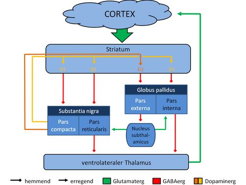
The striosomes receive input from the prefrontal cortex and give outputs to the substantia nigra pars compacta.[17] There are more striosomes present in the dorsal striatum making up 10-15% of the striatal volume, than in the ventral striatum.[16]
Medium spiny neurons (MSNs), which are the principal neurons of the striatum.[2] They are GABAergic and, thus, are classified as inhibitory neurons. Medium spiny projection neurons comprise 95% of the total neuronal population of the human striatum.[2] Medium spiny neurons have two characteristic types: D1-type MSNs and D2-type MSNs.[2][4][18] A subpopulation of MSNs contain both D1-type and D2-type receptors, with approximately40% of striatal MSNs expressing both DRD1 and DRD2mRNA.[2][4][18]
Cholinergicinterneurons release acetylcholine, which has a variety of important effects in the striatum. In humans, other primates, and rodents, these interneurons respond to salient environmental stimuli with stereotyped responses that are temporally aligned with the responses of dopaminergic neurons of the substantia nigra.[19][20] The large aspiny cholinergic interneurons themselves are affected by dopamine through D5 dopamine receptors.[21] Dopamine also directly controls communication between cholinergic interneurons.[22][23]
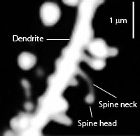
Another well-known afferent is the nigrostriatal connection arising from the neurons of the substantia nigra pars compacta. While cortical axons synapse mainly on spine heads of spiny neurons, nigral axons synapse mainly on spine shafts. In primates, the thalamostriatal afferent comes from the central median-parafascicular complex of the thalamus (see primate basal ganglia system). This afferent is glutamatergic. The participation of truly intralaminar neurons is much more limited. The striatum also receives afferents from other elements of the basal ganglia such as the subthalamic nucleus (glutamatergic) or the external globus pallidus (GABAergic).
Autism spectrum disorder (ASD) is characterized by cognitive inflexibility and poor understanding of social systems. This inflexible behavior originates in defects in the prefrontal cortex as well as the striatal circuits.[62] The defects in the striatum seem to specifically contribute to the motor, social and communication impairments seen in ASD patients. In mice which have an ASD-like phenotype induced via the overexpression of the eukaryotic initiation of translation factor 4E, it has been shown that these defects seem to stem from the reduced ability to store and process information in the striatum, which leads to the difficulty seen in forming new motor patterns, as well as disengaging from existing ones.[63]
Dysfunction
Dysfunction in the ventral striatum can lead to a variety of disorders, most notably depression and obsessive-compulsive disorder. Because of its involvement in reward pathways, the ventral striatum has also been implicated in playing a critical role in addiction. It has been well established that the ventral striatum is strongly involved in mediating the reinforcing effects of drugs, especially stimulants, through dopaminergic stimulation.[64]
The term neostriatum was coined by comparative anatomists comparing the subcortical structures between vertebrates, because it was thought to be a phylogenetically newer section of the corpus striatum. The term is still used by some sources, including Medical Subject Headings.[78]
Other animals
In birds the term used was the paleostriatum augmentatum, while in the new avian terminology listing (as of 2002) for neostriatum this has been changed to the nidopallium.[79]
In non-primate species, the islands of Calleja are included in the ventral striatum.[11]
Striatum highlighted in green on coronal T1 MRI images
Striatum highlighted in green on sagittal T1 MRI images
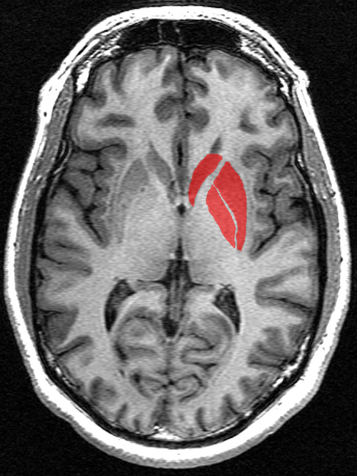
Striatum highlighted in green on transversal T1 MRI images
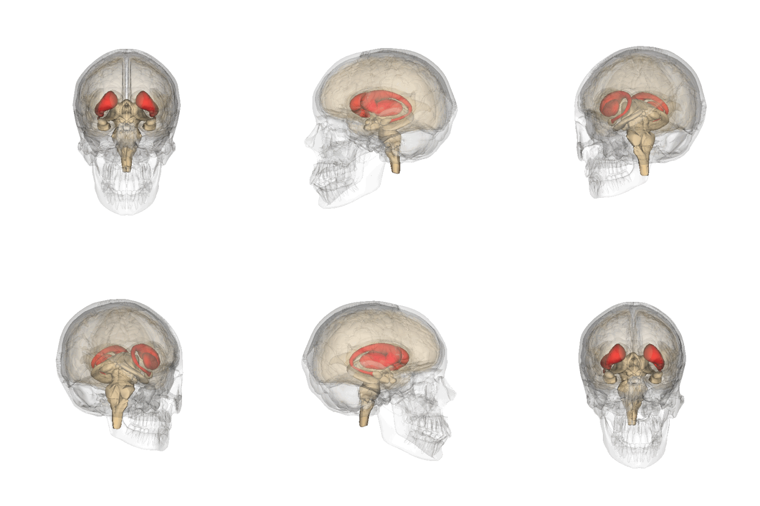
An animation showing the location of the striatum in the human brain.
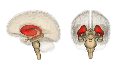
Anatomy of the striatum within the human brain.
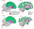
Vocalization subsystems in complex-vocal learners and limited-vocal learners.
Comparative evolution of the striatum and pallium in vertebrates. The ratio of the brain mass devoted to the pallium increases in parallel in various vertebrate taxa.
References
↑"Basal ganglia". BrainInfo. Retrieved 16 August 2015.
12345678910Yager LM, Garcia AF, Wunsch AM, Ferguson SM (August 2015). "The ins and outs of the striatum: Role in drug addiction". Neuroscience. 301: 529–541. doi:10.1016/j.neuroscience.2015.06.033. PMC4523218. PMID26116518. [The striatum] receives dopaminergic inputs from the ventral tegmental area (VTA) and the substantia nigra (SNr) and glutamatergic inputs from several areas, including the cortex, hippocampus, amygdala, and thalamus (Swanson, 1982; Phillipson and Griffiths, 1985; Finch, 1996; Groenewegen et al., 1999; Britt et al., 2012). ... It should also be noted that there is a small population of neurons in the NAc that coexpress both D1 and D2 receptors, though this is largely restricted to the NAc shell (Bertran- Gonzalez et al., 2008).
123456Taylor SB, Lewis CR, Olive MF (February 2013). "The neurocircuitry of illicit psychostimulant addiction: acute and chronic effects in humans". Subst. Abuse Rehabil. 4: 29–43. doi:10.2147/SAR.S39684. PMC3931688. PMID24648786. As described above, the initial rewarding and reinforcing effects of drugs of abuse are mediated by increases in extracellular DA in the NAc shell, and after continued drug use in the NAc core.47,48 After prolonged drug use, drug-associated cues produce increases in extracellular DA levels in the DS and not in the NAc.49
123456789Ferré S, Lluís C, Justinova Z, Quiroz C, Orru M, Navarro G, Canela EI, Franco R, Goldberg SR (June 2010). "Adenosine-cannabinoid receptor interactions. Implications for striatal function". Br. J. Pharmacol. 160 (3): 443–453. doi:10.1111/j.1476-5381.2010.00723.x. PMC2931547. PMID20590556. Two classes of MSNs, which are homogeneously distributed in the striatum, can be differentiated by their output connectivity and their expression of dopamine and adenosine receptors and neuropeptides. In the dorsal striatum (mostly represented by the nucleus caudate-putamen), enkephalinergic MSNs connect the striatum with the external globus pallidus and express the peptide enkephalin and a high density of dopamine D2 and adenosine A2A receptors (they also express adenosine A1 receptors), while dynorphinergic MSNs connect the striatum with the substantia nigra (pars compacta and reticulata) and the entopeduncular nucleus (internal globus pallidus) and express the peptides dynorphin and substance P and dopamine D1 and adenosine A1 but not A2A receptors
↑"striatum | Definition of striatum in English by Oxford Dictionaries". Oxford Dictionaries | English. Archived from the original on 18 January 2018. Retrieved 17 January 2018.
↑Bamford IJ, Bamford NS (October 2019). "The Striatum's Role in Executing Rational and Irrational Economic Behaviors". Neuroscientist. 25 (5): 475–490. doi:10.1177/1073858418824256. PMC6656632. PMID30678530.
↑Báez-Mendoza, Raymundo; Schultz, Wolfram (2013). "The role of the striatum in social behavior". Frontiers in Neuroscience. 7: 233. doi:10.3389/fnins.2013.00233. PMC3857563. PMID24339801.
↑Telford, Ryan; Vattoth, Surjith (February 2014). "MR Anatomy of Deep Brain Nuclei with Special Reference to Specific Diseases and Deep Brain Stimulation Localization". The Neuroradiology Journal. 27 (1): 29–43. doi:10.15274/NRJ-2014-10004. PMC4202840. PMID24571832.
↑"Striatum definition and meaning | Collins English Dictionary". www.collinsdictionary.com.
12Ubeda-Bañon I, Novejarque A, Mohedano-Moriano A, etal. (2007). "Projections from the posterolateral olfactory amygdala to the ventral striatum: neural basis for reinforcing properties of chemical stimuli". BMC Neurosci. 8 103. doi:10.1186/1471-2202-8-103. PMC2216080. PMID18047654.
↑"Ventral Striatum Definition – Medical Dictionary". medicaldictionary.net. Retrieved 18 November 2015.
↑"Ventral Striatum – Medical Definition". www.medilexicon.com. Retrieved 18 November 2015.
↑Turner, Karly M.; Svegborn, Anna; Langguth, Mia; McKenzie, Colin; Robbins, Trevor W. (9 March 2022). "Opposing Roles of the Dorsolateral and Dorsomedial Striatum in the Acquisition of Skilled Action Sequencing in Rats". The Journal of Neuroscience. 42 (10): 2039–2051. doi:10.1523/JNEUROSCI.1907-21.2022. PMC8916752. PMID35086903.
↑Macpherson, Tom; Hikida, Takatoshi (June 2019). "Role of basal ganglia neurocircuitry in the pathology of psychiatric disorders". Psychiatry and Clinical Neurosciences. 73 (6): 289–301. doi:10.1111/pcn.12830. PMID30734985.
12Brimblecombe, K. R.; Cragg, S. J. (2017). "The Striosome and Matrix Compartments of the Striatum: A Path through the Labyrinth from Neurochemistry toward Function". ACS Chemical Neuroscience. 8 (2): 235–242. doi:10.1021/acschemneuro.6b00333. PMID27977131.
123Squire, Larry (2013). Fundamental neuroscience (4.ed.). Amsterdam Heidelberg: Elsevier Academic Press. p.658. ISBN9780123858702.
12Nishi, Akinori; Kuroiwa, Mahomi; Shuto, Takahide (2011). "Mechanisms for the Modulation of Dopamine D1 Receptor Signaling in Striatal Neurons". Frontiers in Neuroanatomy. 5: 43. doi:10.3389/fnana.2011.00043. PMC3140648. PMID21811441.
↑Goldberg, J.A.; Reynolds, J.N.J. (December 2011). "Spontaneous firing and evoked pauses in the tonically active cholinergic interneurons of the striatum". Neuroscience. 198: 27–43. doi:10.1016/j.neuroscience.2011.08.067. PMID21925242.
1 2 Tepper, James M.; Tecuapetla, Fatuel; Koós, Tibor; Ibáñez-Sandoval, Osvaldo (2010). "線条体GABA作動性介在ニューロンの異質性と多様性" . Frontiers in Neuroanatomy . 4 : 150. doi : 10.3389/fnana.2010.00150 . PMC 3016690 . PMID 21228905 .
↑ Koós, Tibor; Tepper, James M. (1999年5月)「GABA作動性介在ニューロンによる線条体投射ニューロンの抑制制御」Nature Neuroscience . 2 (5): 467– 472. doi : 10.1038/8138 . PMID 10321252 .
↑Ibanez-Sandoval, O.; Tecuapetla, F.; Unal, B.; Shah, F.; Koos, T.; Tepper, J. M. (19 May 2010). "Electrophysiological and Morphological Characteristics and Synaptic Connectivity of Tyrosine Hydroxylase-Expressing Neurons in Adult Mouse Striatum". Journal of Neuroscience. 30 (20): 6999–7016. doi:10.1523/JNEUROSCI.5996-09.2010. PMC4447206. PMID20484642.
↑Ibanez-Sandoval, O.; Tecuapetla, F.; Unal, B.; Shah, F.; Koos, T.; Tepper, J. M. (16 November 2011). "A Novel Functionally Distinct Subtype of Striatal Neuropeptide Y Interneuron". Journal of Neuroscience. 31 (46): 16757–16769. doi:10.1523/JNEUROSCI.2628-11.2011. PMC3236391. PMID22090502.
↑English, Daniel F; Ibanez-Sandoval, Osvaldo; Stark, Eran; Tecuapetla, Fatuel; Buzsáki, György; Deisseroth, Karl; Tepper, James M; Koos, Tibor (11 December 2011). "GABAergic circuits mediate the reinforcement-related signals of striatal cholinergic interneurons". Nature Neuroscience. 15 (1): 123–130. doi:10.1038/nn.2984. PMC3245803. PMID22158514.
↑Ernst, Aurélie; Alkass, Kanar; Bernard, Samuel; Salehpour, Mehran; Perl, Shira; Tisdale, John; Possnert, Göran; Druid, Henrik; Frisén, Jonas (February 2014). "Neurogenesis in the Striatum of the Adult Human Brain". Cell. 156 (5): 1072–1083. doi:10.1016/j.cell.2014.01.044. PMID24561062.
↑Inta, D; Lang, U E; Borgwardt, S; Meyer-Lindenberg, A; Gass, P (16 February 2016). "Adult neurogenesis in the human striatum: possible implications for psychiatric disorders". Molecular Psychiatry. 21 (4): 446–447. doi:10.1038/mp.2016.8. PMID26878892.
↑ Kernie, SG; Parent, JM (2010年2月) 「局所虚血性および外傷性脳損傷後の前脳神経新生」 Neurobiology of Disease . 37 (2): 267–74 . doi : 10.1016/j.nbd.2009.11.002 . PMC 2864918. PMID 19909815 .
↑ Rosell, Antonio; Giménez-Amaya, José Manuel (1999年9月)「尾状核への皮質線条体投射の解剖学的再評価:ネコにおける逆行性標識研究」Neuroscience Research . 34 (4): 257– 269. doi : 10.1016/S0168-0102(99)00060-7 . PMID 10576548 .
↑ Stocco, Andrea; Lebiere, Christian; Anderson, John R. (2010). "Conditional Routing of Information to the Cortex: A Model of the Basal Ganglia's Role in Cognitive Coordination" . Psychological Review . 117 (2): 541–74 . doi : 10.1037/a0019077 . PMC 3064519 . PMID 20438237 .
↑ Robbins, Trevor W.; Everitt, Barry J. (1992年4月). "背側および腹側線条体におけるドーパミンの機能". Seminars in Neuroscience . 4 (2): 119–127 . doi : 10.1016/1044-5765(92)90010-Y .
↑Pujol, S.; Cabeen, R.; Sébille, S. B.; Yelnik, J.; François, C.; Fernandez Vidal, S.; Karachi, C.; Zhao, Y.; Cosgrove, G. R.; Jannin, P.; Kikinis, R.; Bardinet, E. (2016). "In vivo Exploration of the Connectivity between the Subthalamic Nucleus and the Globus Pallidus in the Human Brain Using Multi-Fiber Tractography". Frontiers in Neuroanatomy. 10: 119. doi:10.3389/fnana.2016.00119. PMC5243825. PMID28154527.
12Malenka RC, Nestler EJ, Hyman SE (2009). Sydor A, Brown RY (eds.). Molecular Neuropharmacology: A Foundation for Clinical Neuroscience (2nded.). New York: McGraw-Hill Medical. pp.147–148, 321, 367, 376. ISBN978-0-07-148127-4. VTA DA neurons play a critical role in motivation, reward-related behavior (Chapter 15), attention, and multiple forms of memory. This organization of the DA system, wide projection from a limited number of cell bodies, permits coordinated responses to potent new rewards. Thus, acting in diverse terminal fields, dopamine ... helps consolidate multiple forms of memory (amygdala and hippocampus), and encodes new motor programs that will facilitate obtaining this reward in the future (nucleus accumbens core region and dorsal striatum).
↑Kim, BaekSun; Im, Heh-In (2019). "The role of the dorsal striatum in choice impulsivity". Annals of the New York Academy of Sciences. 1451 (1): 92–111. Bibcode:2019NYASA1451...92K. doi:10.1111/nyas.13961. PMID30277562.
↑Jacquemot, Charlotte; Bachoud-Lévi, Anne-Catherine (August 2021). "Striatum and language processing: Where do we stand?". Cognition. 213 104785. doi:10.1016/j.cognition.2021.104785. PMID34059317.
↑Ullman, Michael T.; Corkin, Suzanne; Coppola, Marie; Hickok, Gregory; Growdon, John H.; Koroshetz, Walter J.; Pinker, Steven (March 1997). "A Neural Dissociation within Language: Evidence that the Mental Dictionary Is Part of Declarative Memory, and that Grammatical Rules Are Processed by the Procedural System". Journal of Cognitive Neuroscience. 9 (2): 266–276. doi:10.1162/jocn.1997.9.2.266. PMID23962016.
↑Greengard, P (2001). "The neurobiology of slow synaptic transmission". Science. 294 (5544): 1024–30. Bibcode:2001Sci...294.1024G. doi:10.1126/science.294.5544.1024. PMID11691979.
↑Cachope, R; Cheer (2014). "Local control of striatal dopamine release". Frontiers in Behavioral Neuroscience. 8: 188. doi:10.3389/fnbeh.2014.00188. PMC4033078. PMID24904339.
↑UCL (25 June 2008). "Adventure - it's all in the mind, say UCL neuroscientists". UCL News.
↑Volman, S. F.; Lammel; Margolis; Kim; Richard; Roitman; Lobo (2013). "New insights into the specificity and plasticity of reward and aversion encoding in the mesolimbic system". Journal of Neuroscience. 33 (45): 17569–76. doi:10.1523/JNEUROSCI.3250-13.2013. PMC3818538. PMID24198347.
↑Luna, Beatriz; Sweeney, John A. (June 2004). "The Emergence of Collaborative Brain Function: fMRI Studies of the Development of Response Inhibition". Annals of the New York Academy of Sciences. 1021 (1): 296–309. Bibcode:2004NYASA1021..296L. doi:10.1196/annals.1308.035. PMID15251900.
↑"Department of Physiology, Development and Neuroscience: About the Department". Archived from the original on 11 April 2012. Retrieved 15 December 2007.
↑Choi EY, Yeo BT, Buckner RL (2012). "The organization of the human striatum estimated by intrinsic functional connectivity". Journal of Neurophysiology. 108 (8): 2242–2263. doi:10.1152/jn.00270.2012. PMC3545026. PMID22832566.
↑Steinberg, Laurence (April 2010). "A dual systems model of adolescent risk-taking". Developmental Psychobiology. 52 (3): 216–224. doi:10.1002/dev.20445. PMID20213754.
↑DeLong, Mahlon R.; Wichmann, Thomas (January 2007). "Circuits and Circuit Disorders of the Basal Ganglia". Archives of Neurology. 64 (1): 20–24. doi:10.1001/archneur.64.1.20. PMID17210805.
↑Nestler EJ (December 2013). "Cellular basis of memory for addiction". Dialogues Clin. Neurosci. 15 (4): 431–443. doi:10.31887/DCNS.2013.15.4/enestler. PMC3898681. PMID24459410.
↑Olsen, Christopher M. (December 2011). "Natural rewards, neuroplasticity, and non-drug addictions". Neuropharmacology. 61 (7): 1109–1122. doi:10.1016/j.neuropharm.2011.03.010. PMC3139704. PMID21459101.
↑Simpson, E-H; Kellendonk, C; Kandel, E (11 March 2010). "A Possible Role for the Striatum in the Pathogenesis of the Cognitive Symptoms of Schizophrenia". Neuron. 65 (5): 585–596. doi:10.1016/j.neuron.2010.02.014. PMC4929859. PMID20223196.
↑Fineberg, Naomi A; Potenza, Marc N; Chamberlain, Samuel R; Berlin, Heather A; Menzies, Lara; Bechara, Antoine; Sahakian, Barbara J; Robbins, Trevor W; Bullmore, Edward T; Hollander, Eric (25 November 2009). "Probing Compulsive and Impulsive Behaviors, from Animal Models to Endophenotypes: A Narrative Review". Neuropsychopharmacology. 35 (3): 591–604. doi:10.1038/npp.2009.185. PMC3055606. PMID19940844.
↑Santini, Emanuela; Huynh, Thu N.; MacAskill, Andrew F.; Carter, Adam G.; Pierre, Philippe; Ruggero, Davide; Kaphzan, Hanoch; Klann, Eric (23 December 2012). "Exaggerated translation causes synaptic and behavioural aberrations associated with autism". Nature. 493 (7432): 411–415. Bibcode:2013Natur.493..411S. doi:10.1038/nature11782. PMC3548017. PMID23263185.
↑Everitt, Barry J.; Robbins, Trevor W. (November 2013). "From the ventral to the dorsal striatum: Devolving views of their roles in drug addiction". Neuroscience & Biobehavioral Reviews. 37 (9): 1946–1954. doi:10.1016/j.neubiorev.2013.02.010. PMID23438892.
↑Fedorenko, Evelina (2014). "The role of domain-general cognitive control in language comprehension". Frontiers in Psychology. 5: 335. doi:10.3389/fpsyg.2014.00335. PMC4009428. PMID24803909.
↑Kreisler, A.; Godefroy, O.; Delmaire, C.; Debachy, B.; Leclercq, M.; Pruvo, J.-P.; Leys, D. (14 March 2000). "The anatomy of aphasia revisited". Neurology. 54 (5): 1117–1123. doi:10.1212/wnl.54.5.1117. PMID10720284.
↑ Robles, S G.; Gatignol, P; Capelle, L; Mitchell, MC; Duffau, H (2005年7月) 「言語における優位線条体の役割:術中電気刺激を用いた研究」 Journal of Neurology, Neurosurgery & Psychiatry . 76 (7): 940– 946. doi : 10.1136/jnnp.2004.045948 . PMC 1739710 . PMID 15965199 .
↑ Guenther, Frank H.; Ghosh, Satrajit S.; Tourville, Jason A. (2006年3月1日). 「音節生成の根底にある皮質相互作用の神経モデリングとイメージング」 . Brain and Language . 96 (3): 280–301 . doi : 10.1016/j.bandl.2005.06.001 . PMC 1473986 . PMID 16040108 .