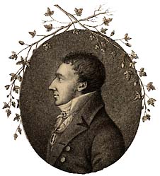
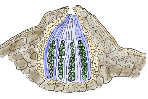
Plural perithecia. A spherical or flask-shaped ascocarp that is sessile or partly immersed in the thallus, with a single opening (ostiole) and enclosed by a distinct wall; a characteristic of pyrenolichens.[345] Although it was in 1831 that Elias Fries first applied the term perithecium to lichen fruit bodies, the word was originally coined by Christiaan Hendrik Persoon in 1794.[346]
Plural phenocortices, phenocortexes. A structure, similar to a cortex, containing hyphal fragments and dead, collapsed algal cells sloughed off from the algal layer.[348]
photobiont
Also defined: phycobiont, cyanobiont. The photosynthetic component of a lichen. This can be either a green alga (known as a phycobiont) or a cyanobacteria (known as a cyanobiont).[349] The term "phycobiont" was proposed by George Scott in 1957.[5]
A historical lichenological term with multiple meanings. Originally introduced by Fries (1831) as "Phyco-Lichenes" for lichens morphologically resembling seaweeds. Later redefined by Massalongo (1855) for homoiomerous cyanobacterial lichens, then by Diels (1936) for lichens with Phycomycetes as their fungal partner. More recently proposed but discouraged as a term for green algal lichens, with "chlorolichen" recommended instead as a counterpart to "cyanolichen". Generally considered obsolete in modern lichenology due to its ambiguous history.[98]
phyllidium
Plural phyllidia. A small leaf-like or scale-like propagule that is corticate and has distinct upper and lower sides (i.e., it is dorsiventral); it originates from the margins or on the upper surface of thallus.[353] Phyllidia occur in some species of the Lecanorales and the Peltigerales.[354]
Characterised by longitudinal folds forming pleats, often used to describe closely adjacent, markedly convex thallus lobes or elongated areoles that display a "folded" appearance.[362]
plurilocular
Also multilocular. Having many cavities or locules; used to describe spore structure.[363]
A lichen test performed by shining a polarized light at a lichen structure in microscopic view; in the genus Hypogymnia, the presence (POL+) or absence (POL−) of POL-sensitive crystals in the hypothecium is a useful character to help distinguish species.[364]
polarilocular
Also placodiomorphic,[256]polar-diblastic, polaridiblastic, polaribilocular, polocellate. A spore divided into two components (locules) separated by a central septum with a perforation or isthmus.[365][366] The term was first used by Wilhelm Körber in 1855 (as "polari-dyblastae") to describe the spores of Rhizocarpon and Umbilicaria. It was anglicized to "polari-bilocular" by William Mudd in 1861, and finally shortened to polarilocular by the Henri Olivier in 1882.[4]
A morphotype of corticolousthelotremoid lichens used to describe characteristics of apothecial and thallus morphology. Praestantoid lichens have large and prominent apothecia with small pores; pores with "finger" (columella); black walls (viewed in microscopic section); and a smooth, more or less shiny thallus. This morphotype occurs in the praestansgroup of the genus Ocellularia.[30]
Sticking out from the surface of the thallus.[370] A more precise definition has been suggested by Aptroot and Lücking, who propose that the term applies to ascomata and pseudostromata that are more than 1/2 above the level of the thallus, and have a base that is expanded outwards.[183]
proper margin
Also proper exciple, true exciple, ectal excipulum, medullary excipulum, proprium. A ring of tissue around the disk of a lecanorineapothecium; this tissue, which originates from the medulla, is not lichenized, and is internal to the thalline margin (if present).[371] The term "proper margin" was first used by Erik Acharius in 1803; in 1825 Elias Fries changed the noun and called it "proper exciple".[3]
Plural prosenchymata. A type of plectenchyma in which the constituent fungal hyphae are arranged parallel to each other, such that individual hyphae can be clearly distinguished using microscopy.[372]
prosoplectenchyma
Plural prosoplectenchymata. A type of plectenchyma, common in the thalluscortex of lichens, in which the constituent fungal hyphae are aligned in a particular direction.[372]
A fungal layer upon which an algae-containing thallus may develop, lacking photobiont; usually white, brown, or black, and found between the areoles and at the growing margins of crustose lichens.[373] The term was first used by Georg Meyer in 1825.[3]
prototunicate
A form of unitunicateascus in which the wall breaks down before maturity (thus releasing its ascospores), and which lacks differentiated apical structures.[374]
proximal
Positioned close to a point of origin or near the center of a body.[375]
pruina
A powdery, frost-like or flour-like deposit on a surface. In lichens, pruina is often the result of the accumulation of crystalline hydrates of calcium oxalate, of lichen products, or sometimes of the dead or dying cells of the epinecral layer.[376]
Also pseud-. A prefix meaning "false";[377] used in terminology to denote something is false, or that one structure resembles something else, such as the pseudocyphella resembling the cyphella.[375]
Plural pseudocortices, pseudocortexes. A boundary layer of the thallus containing distinct hyphae that are not organized into a regular tissue structure;[378] sometimes used to refer to the false cortex present on the outer layer of pseudopodetia, such as those found in the lichen Pycnothelia papillaria.[379]
Plural pseudocyphellae. Small openings in the cortex of a lichen, where the medulla is exposed to air, and there are no specialized cells surrounding the cavity.[380] The term was first used by William Nylander in 1858.[4]
Also cataphysis. A paraphysis-like hypha that forms in the locule or perithecial cavity before the formation of the ascus; it grows downward from the top of the cavity to the base of the ascomata.[382]
A lichen with flask-shaped fruiting bodies (perithecia) that develop from the fungal partner. Originally thought to form a natural group, molecular studies have shown pyrenocarpous lichens to be highly polyphyletic, evolving independently in multiple fungal lineages. Most belong to Chaetothyriomycetidae (e.g., Verrucariales, Pyrenulales), with some in Dothideomycetes (e.g., Arthopyreniaceae, Trypetheliaceae) and others unusually placed in Lecanoromycetes (e.g., Porinaceae, Protothelenellaceae, Thelenellaceae). Pyrenocarpous lichens are notably absent from the classical pyrenomycete class Sordariomycetes. This diverse group demonstrates that ascoma morphology alone is insufficient for determining evolutionary relationships, with convergent evolution of perithecial fruiting bodies occurring multiple times across fungal lineages.[393]
Also recurvate, reflexed.[398] Curved or bent back; in lichens, these terms are used to describe the tips of branches or lobes that are curved up or down, or back onto themselves.[399]
redingerioid
A morphotype of corticolousthelotremoid lichens used to describe characteristics of apothecial and thallus morphology. Redingerioid lichens have immersed apothecia with linear slit; slit filled with irregular structures; black walls (viewed in microscopic section); and a smooth, more or less shiny thallus. This morphotype occurs in the genera Redingeria and Stegobolus.[30]
reimnitzioid
A morphotype of corticolousthelotremoid lichens used to describe characteristics of apothecial and thallus morphology. Reimnitzioid lichens have open apothecia with erect lobules, and a rough thallus containing crystals; this morphotype occurs in the genus Reimnitzia.[30]
A single hyphal strand on the thallus underside that serves as an attachment organ.[403]
rhodostromoid
A morphotype of corticolousthelotremoid lichens used to describe characteristics of apothecial and thallus morphology. Rhodostromoid lichens have large and prominent apothecia with small pores; pore with "finger" (columella); black walls and pigment (viewed in microscopic section); and a smooth, more or less shiny thallus. This morphotype occurs in the rhodostromagroup of the genus Ocellularia.[30]
A morphotype of corticolousthelotremoid lichens used to describe characteristics of apothecial and thallus morphology. Schizotremoid lichens lack apothecia and have schizidia, and a smooth, more or less shiny thallus; this morphotype occurs in the genus Stegobolus.[30]
Plural scleroplectenchymata. A type of plectenchyma comprising thick-walled hyphae that are stuck closely together; present as a component of the stereoma tissue supporting the thallus in the genera Cladonia and Alectoria.[415]
scrobiculate
Having large, shallow depressions that are narrowly separated by rounded ridges.[195]Compare: faveolate, foveolate.
Lacking a stem.[421] A more precise definition has been suggested by Aptroot and Lücking, who propose that the term applies to ascomata and pseudostromata that are more than 3/4 above the level of the thallus, with a constricted base.[183]
seta
A stiff hair or bristle-like structure, typically thick-walled and sharp-pointed, found on some lichens.[422]
sibling species
Closely related species that are morphologically indistinguishable but can be distinguished by non-morphological traits, such as chemistry or genetic differences. Initially defined as species recognized mainly through cryptic or non-morphological discontinuities, the concept has evolved to specifically refer to cryptic species that form a monophyletic group, meaning they share a common ancestor not shared with any other species. This concept is a subset of the broader terms species pair and "cryptic species".[423]
sigmoid
Curved upon itself twice, like the letter "S".[424]
simple
Lacking branches or divisions; in lichenology, it is used to describe structures such as thalli or rhizines, or spores without septa.[425]
sinuose
Also sinuous. Wavy, winding, or having a serpentine form, with alternating curves and indentations along the margin or edge.[426]
Plural soralia. A part of the thallus where the cortex has cracked or broken down and soredia are produced. Soralia can be further characterized as diffuse if they are spread out on the upper thallus surface as a continuous layer, or delimited if they are confined to a more restricted area. If soralia originate in tubercules they are tuberculate, while they are fissural if they are created in fissures.[429] The term was proposed by Johannes Reinke in 1895.[5]
Plural soredia. A powdery to granular reproductive propagule that is not covered with a well-defined cortex (in contrast to isidia, and contains both algal (photobiont) and fungal (mycobiont) components.[429] The term was first used by Erik Acharius in 1803.[3]
Two lichen species that are identical morphologically, anatomically, and chemically, but can be distinguished by their sexual versus asexual reproductive strategies; the fertile taxon is known as the primary species, while the vegetatively reproducing taxon is known as the secondary species.[369] The use of molecular methods to analyze putative species pairs has shown that the underlying phylogenetic situation is more complex than had been assumed, and not necessarily correlated with reproductive strategy.[431]See related: sibling species.
spermogonium
Also spermagone, spermagonium. In lichenology, an obsolete term for pycnidium.[432]
A small spine; in some fruticose lichens of the Lecanoromycetes, it refers to a small cylindrical outgrowth, with a narrow base, in which the central axis is not connected with the central axis of the main branch.[433]
spinulose
Also spinulous. Covered with or having small spines (spinules) or spiny projections.[433]
A lichen species that is extremely similar (or identical) in external morphology, anatomy, chemistry, and spore size to another, but that is placed in a different genus solely based on differences in spore septation and/or spore colour. The term was introduced by Michael Wirth and Mason Hale in their 1978 monograph about the Graphidaceae, a family in which sporomorphs are common.[437]
A spot analysis used to help identify lichens; it is performed by placing a drop of a reagent on different parts of the lichen and noting any color change associated with application of the reagent. The four most common tests are C, K, KC, and PD.[439]
squamule
A small, flattened, scale-like structure that forms part of the thallus of certain lichens.[440]
Brush-like, with many short, more or less perpendicular lateral branches. In lichenology, used to refer to rhizine structure.[441]
stegoboloid
A morphotype of corticolousthelotremoid lichens used to describe characteristics of apothecial and thallus morphology. Stegoboloid lichens have prominent apothecia with wide pores; pore filled with irregular structures; black walls (viewed in microscopic section); and a smooth, more or less shiny thallus. This morphotype occurs in the genus Stegobolus.[30]
Plural stromata. A dense mass of vegetativehyphae that supports spore-bearing structures.[443] In lichens, the stroma is often hard and carbonaceous.[403]
sub-
A prefix meaning "below", "under", "somewhat, or "almost".[444] Also used in front of names of taxonomic ranks to indicate intermediate categories, e.g. subspecies or subgenus.[445]
Also subicule.Plural subicula. A layer of loosely-compacted mycelia that covers the substrate and cushions fruiting bodies such as apothecia and perithecia.[446] The texture of the subiculum can be described as net-like, wool-like, or crust-like.[444]
substrate
Also substratum; plural substrata. The surface or base upon which a lichen grows or is attached. Although the terms substratum and substrate are often used equivalently in lichenology, the latter term has different meanings in microbiology and in enzymology.[447]
subulate
Slender and narrowing to a fine point; awl-shaped.[448]
A morphotype of corticolousthelotremoid lichens used to describe characteristics of apothecial and thallus morphology. Tenuitremoid lichens have immersed apothecia with small pores and a black margin; pore with "finger" (columella); black walls (viewed in microscopic section); and a smooth, more or less shiny thallus. This morphotype occurs in the genus Clandestinotrema.[30]
tegulicolous lichen
A lichen that lives on tiles.[450] In general, these are calcicolous lichens or lichens that are indifferent as to their substrate.[451]
Describing a cylindrical or rod-like structure that is round in cross-section.[428]
teretiform
Describing a shape that is nearly cylindrical or rod-like, similar to but not exactly terete; circular in cross-section, gradually tapering towards one end.[453]
Also thalline exciple, excipulum thallinum. A rim of tissue around the disk of a lecanorineapothecium; this tissue, external to the inner proper margin, is made of tissue with a structure similar to that of the thallus.[455] The term "thalline margin" was first used by Erik Acharius in 1803; in 1825 Elias Fries changed the noun and called it "thalline exciple".[3]
Plural thalloconidia. A dark brown, smooth to wrinkled propagule arising directly from a thallus, particularly the lower cortex and/or the rhizines. They are found in some species of Umbilicaria, and similar structures arise from the prothallus of some species in the genera Protoparmelia, Rhizoplaca, and Sporastatia. Thalloconidia have distinct cell layers in their walls, and comprise between 1 and about 2500 cells.[457][458]
Plural thalli. The body of a lichen,[460] made up of both fungal and algal or cyanobacterial cells.[461] The term was first used by Erik Acharius in 1803.[3]
A morphological group of lichens within the Graphidaceae, the largest family of crustose lichens.[463] Thelotremoid lichens are characterized by immersed-erumpent, rounded ascomata, non-branched to slightly branched paraphyses, mostly distoseptate ascospores, and mostly a prosoplectenchymatousexcipulum. Thelotremataceae, a traditional family of lichens, has been included in Graphidaceae, and its species are now informally accepted as thelotremoid lichens.[463]
tholus
Plural tholi.Also dome. The apical, often thickened part of the inner wall in a bitunicate ascus.[464]
tomentum
Plural tomenta. Also defined: tomentose. A layer of short interwoven or coiled fungal hyphae with a texture similar to velvet. In lichens, the tomentum projects from the lower cortex and serves to help it attach to its substrate. Structures with this type of hyphae are called tomentose.[465] Tomentose surfaces are found in foliose genera such as Lobaria, Pseudocyphellaria, and Sticta.[466]
topeliopsidoid
A morphotype of corticolousthelotremoid lichens used to describe characteristics of apothecial and thallus structure. It pertains to lichens where the apothecia are either prominent or sessile, often hidden between the substratum, and they open with multiple, typically regular "teeth" that stay relatively curved over the hardly visible disk. The margins tend to peel off, meaning the overlaying thallus cortex separates from the underlying marginal thallus tissue, but no distinct, clean split between thallus margin and excipulum is formed. This morphotype is seen in species like Chapsa meridensis and Topeliopsis.[467]
trabecula
Plural trabeculae. In the genus Umbilicaria, they are rib- or strap-shaped structures radiating outward from the umbilicus that merge towards the mid-zone of the thallus undersurface.[276]
trentepohlioid
Also trentepohlialean.[468] Resembling or belonging to the green algal genus Trentepohlia; trentepohlioid cells are filamentous (elongated and cylindrical), multicellular, and have a yellow to orange colour.[469]
Also defined: funiculus, umbilicus, umbilical cord. A lichen with a concave, circular, leafy thallus that is joined to its substrate only by its central part, called an umbilicus, umbilical cord or funiculus.[472][473]
A type of ascus with a single functional layer; the rigid internal and external wall layers do not separate during release of the ascospores. Most ascomycetes have unitunicate asci.[477]
Deeply cup-shaped or urn-shaped; in lichens, the term is used to describe some apothecia with a sunken hymenium and elevated parathecium that forms a narrow mouth.[479]
A lichen test performed by shining a long-wavelength ultraviolet light (350nm) at a lichen structure to check if it fluoresces; a positive test (abbreviated as UV+) indicates the presence of certain lichen products. Xanthone compounds in the cortex tend to fluoresce yellow, orange, or red, while depsides and depsidones in the medulla fluoresce blue to white.[481]
A lichen not attached to a substrate, typically able to be blown around by wind.[482]
vegetative
Also assimilative. Having to do with the growth phase of an organism before reproduction, including spore germination, growth, development and asexual multiplication.[483]
"Vitricolous lichens – Lichen website". Australian National Botanic Gardens - Botanical Web Portal. Australian National Herbarium. 20 October 2009. Retrieved 18 October 2022.
"Definition of mycobiont". Collins English Dictionary. 2022a. Retrieved 23 July 2022.
"Definition of phycobiont". Collins English Dictionary. 2022b. Retrieved 23 July 2022.
Ahti, T. (1982). "The morphological interpretation of cladoniiform thalli in lichens". The Lichenologist. 14 (2): 105–113. doi:10.1017/s0024282982000255.
Aptroot, André; Lücking, Robert (2016). "A revisionary synopsis of the Trypetheliaceae (Ascomycota: Trypetheliales)". The Lichenologist. 48 (6): 763–982. doi:10.1017/s0024282916000487.
Breuss, Othmar (2010). "An updated world-wide key to the catapyrenioid lichens (Verrucariaceae)". Herzogia. 23 (2): 205–216. doi:10.13158/heia.23.2.2010.205.
Brodo, Irwin M.; Sharnoff, Sylvia Duran; Sharnoff, Stephen (2001). Lichens of North America. New Haven, Connecticut: Yale University Press. ISBN0-300-08249-5.
Brodo, Irwin (2016). "Glossary". Keys to Lichens of North America: Revised and Expanded. New Haven, Connecticut: Yale University Press. pp.369–382. ISBN978-0-300-19573-6.
Cannon, Paul F.; Kirk, Paul M. (2007). Fungal Families of the World. Wallingford: CABI. ISBN978-0-85199-827-5. OCLC60741230.
Diederich, Paul; Lawrey, James D.; Ertz, Damien (2018). "The 2018 classification and checklist of lichenicolous fungi, with 2000 non-lichenized, obligately lichenicolous taxa". The Bryologist. 121 (3): 340–425. doi:10.1639/0007-2745-121.3.340.
Dobson, Frank S. (2011). Lichens: An Illustrated Guide to the British and Irish Species. Slough, UK: Richmond Publishing. ISBN978-0-85546-315-1.
Ferraro, Lidia Itatí (2004). "Morphological diversity in the hyphopores of Gomphillaceae (Ostropales, lichenized Ascomycetes)"(PDF). Fungal Diversity. 15: 153–169.
Fjelde, Markus Osaland; Timdal, Einar; Haugan, Reidar; Bendiksby, Mika (2024). "Paraphyly and cryptic diversity unveils unexpected challenges in the "naked lichens" (Calvitimela, Lecanoromycetes, Ascomycota)". Molecular Phylogenetics and Evolution. 190 107944. doi:10.1016/j.ympev.2023.107944. hdl:10852/106762.
Fraser, Robert H.; Pouliot, Darren; van der Sluijs, Jurjen (2021). "UAV and high resolution satellite mapping of forage lichen (Cladonia spp.) in a rocky Canadian Shield landscape". Canadian Journal of Remote Sensing. 48 (1): 5–18. doi:10.1080/07038992.2021.1908118.
Garrido-Benavent, Isaac; Pérez-Ortega, Sergio (2017). "Past, present, and future research in bipolar lichen-forming fungi and their photobionts". American Journal of Botany. 104 (11): 1660–1674. doi:10.3732/ajb.1700182.
"Glossary of Terms". British Lichen Society. 2022. Retrieved 23 July 2022.
Goward, Trevor (1986). "Brodoa, a new lichen genus in the Parmeliaceae". The Bryologist. 89 (3): 219–223. doi:10.2307/3243288. JSTOR3243288.
Grube, Martin; Hawksworth, David L. (2007). "Trouble with lichen: the re-evaluation and re-interpretation of thallus form and fruit body types in the molecular era". Mycological Research. 111 (9): 1116–1132. doi:10.1016/j.mycres.2007.04.008. PMID17698333.
Hafellner, Josef; Kalb, Klaus (1995). "Studies in Trichotheliales ordo novus". Studies in Lichenology with Emphasis on Chemotaxonomy, Geography and Phytochemistry. Festschrift Ch. Leuckert. Bibliothecia Lichenologica. Vol.57. Berlin-Stuttgart: J. Cramer in der Gebrüder Borntraeger Verlagsbuchhandlung. pp.161–186. ISBN978-3-44-358036-0.
Hawksworth, D. L. (1988). "The variety of fungal-algal symbioses, their evolutionary significance, and the nature of lichens". Botanical Journal of the Linnean Society. 96 (1): 3–20. doi:10.1111/j.1095-8339.1988.tb00623.x.
Hertel, Hannes (2009). "A new key to cryptothalline species of the genus Lecidea (Lecanorales)". In Aptroot, A.; Seaward, M.R.D.; Sparrius, L.B. (eds.). Biodiversity and Ecology of Lichens – Liber Amicorum Harrie Sipman. Bibliotheca Lichenologica. Vol.99. Berlin/Stuttgart: J.Cramer in der Gebrüder Borntraeger Verlagsbuchhandlung. pp.185–204. ISBN978-3-443-58078-0.
Honegger, Rosmarie (1991). "Functional aspects of the lichen symbiosis". Annual Review of Plant Physiology and Plant Molecular Biology. 42 (1): 553–578. doi:10.1146/annurev.pp.42.060191.003005.
Jagtap, Vineeta; Tripathi, Manish; Joshi, Yogesh (2013). "First report on the occurrence of plasticolous lichens from Uttarakhand, India"(PDF). Journal of Applied and Natural Science. 5 (2): 342–344. doi:10.31018/jans.v5i2.329.
Kantvilas, G. & Jarman, S.J. (1999). Lichens of rainforest in Tasmania. Canberra, ACT, Australia: Australian Biological Resources Study. ISBN0-642-56802-2.
Kantvilas, Gintaras (2023). "Three new species of siphuloid lichens, with a first key to the genus Parasiphula". The Lichenologist. 55 (1): 17–25. doi:10.1017/s0024282922000421.
Kitaura, Marcos J.; Marcelli, Marcelo P. (2013). "A revision of Leptogium species with spherical-celled hairs (section Mallotium p.p.)". The Bryologist. 116 (1): 15–27. doi:10.1639/0007-2745-116.1.015.
Kirk, Paul M.; Cannon, Paul F.; Minter, David W.; Stalpers, Joost A., eds. (2008). Dictionary of the Fungi. Vol.10. CAB International. ISBN978-1-84593-933-5.
Kistenich, Sonja; Timdal, Einar; Bendiksby, Mika; Ekman, Stefan (2018). "Molecular systematics and character evolution in the lichen family Ramalinaceae (Ascomycota: Lecanorales)". Taxon. 67 (5): 871–904. doi:10.12705/675.1. hdl:11250/2599544.
Korf, Richard P. (1962). "A synopsis of the Hemiphacidiaceae, a family of the Helotiales (Discomycetes) causing needle-blights of conifers". Mycotaxon. 54 (1): 12–33.
Krzewicka, Beata (2004). "The lichen genera Lasallia and Umbilicaria in the Polish Tatra mts". Polish Botanical Studies. 17: 1–88.
Lange, Otto L.; Wagenitz, Gerhard (2003). "What is a 'phycolichen'? Differences and changes in the meaning of an old lichenological term". The Lichenologist. 35 (4): 341–345. doi:10.1016/S0024-2829(03)00038-0.
Laundon, Jack R. (1995). "On the classification of lichen photomorphs". Taxon. 44 (3): 387–389. doi:10.2307/1223409. JSTOR1223409.
Lendemer, James C.; Buck, William R.; Harris, Richard C. (September 2016). "Two new host-specific hepaticolous species of Catinaria (Ramalinaceae)". The Lichenologist. 48 (5): 441–449. doi:10.1017/S0024282916000438.
Lücking, Robert; Leavitt, Steven D.; Hawksworth, David L. (2021). "Species in lichen-forming fungi: balancing between conceptual and practical considerations, and between phenotype and phylogenomics". Fungal Diversity. 109 (1): 99–154. doi:10.1007/s13225-021-00477-7.
"Lichen Basics". North American Mycological Association. Retrieved 15 October 2022.
Lumbsch, H. Thorsten; Huhndorf, Sabine M. (2007). "Whatever happened to the pyrenomycetes and loculoascomycetes?". Mycological Research. 111 (9): 1064–1074. doi:10.1016/j.mycres.2007.04.004. PMID18029164.
MacKenzie Lamb, I. (1968). "Appendix II. Glossary of Technical Terms". Antarctic Lichens. II. The Genera Buellia and Rinodina (Report). British Antarctic Survey Scientific Reports. British Antarctic Survey.
McCune, B.; Wang, L.S. (2014). "The lichen genus Hypogymnia in southwest China". Mycosphere. 5 (1): 27–76. doi:10.5943/mycosphere/5/1/2.
Meyer, Barbara; Printzen, Christian (2000). "Proposal for a standardized nomenclature and characterization of insoluble lichen pigments". The Lichenologist. 32 (6): 571–583. doi:10.1006/lich.2000.0294.
Mitchell, M.E. (2014). "De Bary's legacy: the emergence of differing perspectives on lichen symbiosis"(PDF). Huntia. 15 (1): 5–22.
Moncada, Bibiana; Lücking, Robert; Betancourt-Macuase, Luisa (2013). "Phylogeny of the Lobariaceae (lichenized Ascomycota: Peltigerales), with a reappraisal of the genus Lobariella". The Lichenologist. 45 (2): 203–263. doi:10.1017/s0024282912000825.
Moncada, Bibiana; Rincón-Murillo, Diego; Lücking, Robert (2023). "Three new lobarioid lichens (lichenized Ascomycota: Peltigeraceae) from Colombia, honoring Enrique Forero". Revista de la Academia Colombiana de Ciencias Exactas, Físicas y Naturales. 47 (184): 619–640. doi:10.18257/raccefyn.1978.
Nelsen, Matthew P.; Plata, Eimy Rivas; Andrew, Carrie J.; Lücking, Robert; Lumbsch, H. Thorsten (2011). "Phylogenetic diversity of trentepohlialean algae associated with lichen-forming fungi". Journal of Phycology. 47 (2): 282–290. doi:10.1111/j.1529-8817.2011.00962.x. PMID27021860.
Orange, A.; James, P.W.; White, F.J. (2001). Microchemical Methods for the Identification of Lichens. British Lichen Society. ISBN978-0-9540418-0-9.
Øvstedal, D.O.; Lewis Smith, R.I. (2001). "Glossary". Lichens of Antarctica and South Georgia. A Guide to Their Identification and Ecology. Cambridge, UK: Cambridge University Press. pp.367–374. ISBN978-0-521-66241-3.
Poelt, J. (1980). "Physcia opuntiella spec. nov. und die Lebeusform der Sprosseuden Flechten" [Physcia opuntiella spec. nov. and the life form of the branching lichens]. Flora. 169 (1): 23–31. doi:10.1016/s0367-2530(17)31162-3.
Poelt, J.; Obermayer, W. (1990). "Über Thallosporen bei einigen Krustenflechten"[On thallospores in some crustose lichens]. Herzogia (in German). 8 (3–4): 273–288. doi:10.1127/herzogia/8/1990/273.
Rikkinen, Jouko (April 2015). "Cyanolichens". Biodiversity and Conservation. 24 (4): 973–993. doi:10.1007/s10531-015-0906-8.
Nash III, Thomas H., ed. (2008). Lichen Biology (2nded.). Cambridge, UK: Cambridge University Press. ISBN978-0-521-69216-8.
Nash, T.H. III (2008). "1. Introduction". In Nash III, Thomas H. (ed.). Lichen Biology (2nded.). Cambridge, UK: Cambridge University Press. pp.1–8. ISBN978-0-521-69216-8.
Honegger, R. (2008). "3.Mycobionts". In Nash III, Thomas H. (ed.). Lichen Biology (2nded.). Cambridge, UK: Cambridge University Press. pp.27–39. ISBN978-0-521-69216-8.
Büdel, B.; Scheidegger, C. (2008). "4.Thallus morphology and anatomy". In Nash III, Thomas H. (ed.). Lichen Biology (2nded.). Cambridge, UK: Cambridge University Press. pp.40–68. ISBN978-0-521-69216-8.
Nelsen, Matthew P.; Lücking, Robert; Aptroot, André; Andrew, Carrie J.; Cáceres, Marcela; Plata, Eimy Rivas; Gueidan, Cécile; da Silva Canêz, Luciana; Knight, Allison; Ludwig, Lars R.; Mercado-Díaz, Joel A.; Parnmen, Sittiporn; Lumbsch, H. Thorsten (2014). "Elucidating phylogenetic relationships and genus-level classification within the fungal family Trypetheliaceae (Ascomycota: Dothideomycetes)". Taxon. 63 (5): 974–992. doi:10.12705/635.9.
Nelsen, Matthew P.; Lücking, Robert; Boyce, C. Kevin; Lumbsch, H. Thorsten; Ree, Richard H. (2020). "The macroevolutionary dynamics of symbiotic and phenotypic diversification in lichens". Proceedings of the National Academy of Sciences. 117 (35): 21495–21503. doi:10.1073/pnas.2001913117. PMC7474681. PMID32796103.
Papong, Khwanruan; Boonpragob, Kansri; Mangold, Armin; Divakar, Pradeep K.; Lumbsch, H. Thorsten (2010). "Thelotremoid lichen species recently described from Thailand: a re-evaluation". The Lichenologist. 42 (2): 131–137. doi:10.1017/s0024282909990405.
Randlane, Tiina; Tõrra, Tiiu; Saag, Andres; Saag, Lauri (2009). "Key to European Usnea species". In Thell, Arne; Seaward, Mark R. D.; Feuerer, Tassilo (eds.). Diversity of Lichenology – Anniversary Volume. Bibliotheca Lichenologica. Vol.100. Stuttgart: J.Kramer. pp.419–462. ISBN978-3-443-58079-7.
Rapai, Sean B.; McMullin, Richard Troy; Newmaster, Steven G. (2012). "The importance of macrolichen traits and phylogeny in forest community assemblage along a high elevation gradient in southwestern British Columbia". Forest Ecology and Management. 274: 231–240. doi:10.1016/j.foreco.2012.02.018.
Rivas Plata, Eimy; Lücking, Robert; Sipman, Harrie J. M.; Mangold, Armin; Kalb, Klaus; Lumbsch, H. Thorsten (2010). "A world-wide key to the thelotremoid Graphidaceae, excluding the Ocellularia-Myriotrema-Stegobolus clade". The Lichenologist. 42 (2): 139–185. doi:10.1017/s0024282909990491.
Sanders, William B. (2004). "Bacteria, algae, and phycobionts: maintaining useful concepts and terminology". The Lichenologist. 36 (5): 269–275. doi:10.1017/s0024282904014343.
Sérusiaux, E.; Lücking, R. (2007). "Gallaicolichen, a new genus of foliicolous lichen with unique diaspores". In Kärnefelt, Ingvar; Thell, Arne (eds.). Lichenological Contributions in Honour of David Galloway. Bibliotheca Lichenologica. Vol.95. Berlin/Stuttgart: J. Cramer. pp.509–516. ISBN978-3-443-58074-2.
Smith, C.W.; Aptroot, A.; Coppins, B.J.; Fletcher, A.; Gilbert, O.L.; James, P.W.; Wolseley, P.A., eds. (2009). "Glossary". The Lichens of Great Britain and Ireland. London: British Lichen Society. pp.21–39.
Suryanarayanan, Trichur S.; Thirunavukkarasu, Nagamani (2017). "Endolichenic fungi: the lesser known fungal associates of lichens". Mycology. 8 (3): 189–196. doi:10.1080/21501203.2017.1352048. PMC6059131. PMID30123639.
Upreti, Dalip K.; Rai, Himanshu, eds. (2013). Terricolous Lichens in India. Vol.1: Diversity Patterns and Distribution Ecology. New York: Springer. ISBN978-1-4614-8736-4.
Silverstein, Alvin; Silverstein, Virginia B.; Silverstein, Robert A. (1996). Fungi. New York: Twenty-First Century Books. ISBN0-8050-3520-6.
Smith, Annie Lorrain (1921). Lichens. London: Cambridge University Press. pp.1–504.
Thell, Arne; Crespo, Ana; Divakar, Pradeep K.; Kärnefelt, Ingvar; Leavitt, Steven D.; Lumbsch, H. Thorsten; Seaward, Mark R.D. (2012). "A review of the lichen family Parmeliaceae – history, phylogeny and current taxonomy". Nordic Journal of Botany. 30 (6): 641–664. doi:10.1111/j.1756-1051.2012.00008.x.
Thüs, Holger; Schultz, Matthias (2009). "Glossary". In Büdel, Burkhard; Gärtner, Georg; Krienitz, Lothar; Preisig, Hans R.; Schagerl, Michael (eds.). Fungi. Part 1: Lichens. Süßwasserflora von Mitteleuropa [Freshwater Flora of Central Europe]. Vol.21/1. Heidelberg: Spektrum Akademischer Verlag. pp.14–23. doi:10.1007/978-3-8274-2299-6_2. ISBN978-3-8274-1594-3.
Tripp, Erin (2017). Field Guide to the Lichens of White Rocks (Boulder, Colorado). University Press of Colorado. ISBN978-1-60732-554-3.
Ulloa, Miguel; Hanlin, Richard T. (2012). Illustrated Dictionary of Mycology (2nded.). St.Paul, Minnesota: The American Phytopathological Society. ISBN978-0-89054-400-6.
Vondrák, Jan; Svoboda, Stanislav; Zíbarová, Lucie; Štenclová, Lenka; Mareš, Jan; Pouska, Václav; Košnar, Jiří; Kubásek, Jiří (2023). "Alcobiosis, an algal-fungal association on the threshold of lichenisation". Scientific Reports. 13 (2957): 2957. doi:10.1038/s41598-023-29384-4. PMC9975235. PMID36854763.
Wirth, M.; Hale Jr., M.E. (1978). Morden-Smithsonian Expedition to Dominica: The Lichens (Graphidaceae). Smithsonian Contributions to Botany. Vol.40. pp.1–64.
Categories:
Lists of lichens
Glossaries of biology
Hidden categories:
Articles with short description
Short description with empty Wikidata description
Use American English from November 2022
All Wikipedia articles written in American English
Pages using multiple image with auto scaled images