
Loading article…
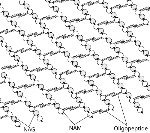
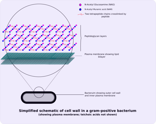
ペプチドグリカン、ムレイン、またはムコペプチドは、細菌の細胞質膜を囲む網目状の層(サッキュラス)を形成する糖とアミノ酸からなる多糖類である独特な巨大高分子です。 [ 1 ]糖成分は、β-(1,4)結合したN-アセチルグルコサミン(NAG)とN-アセチルムラミン酸(NAM)の交互に並んだ残基から構成されています。N-アセチルムラミン酸には、 3 ~5個のアミノ酸からなるオリゴペプチド鎖が結合しています。ペプチド鎖は、別の鎖のペプチド鎖と架橋結合して3D網目状の層を形成することができます。[ 1 ] [ 2 ]ペプチドグリカンは、細菌の細胞壁において構造的な役割を果たし、構造的な強度を与えるとともに、細胞質の浸透圧に対抗します。この繰り返し結合により、細胞形態の維持と高浸透圧への耐性に不可欠な高密度のペプチドグリカン層が形成され、これは定期的にペプチドグリカンの生成によって置き換えられます。ペプチドグリカンの加水分解と合成は、細胞が成長および増殖するために必ず起こる2つのプロセスであり、この技術は、現在の物質の切断、新しい物質の挿入、および既存の物質と新しい物質の再架橋という3つの段階で実行されます。[ 3 ]
ペプチドグリカン層は、グラム陽性菌(20~80ナノメートル)ではグラム陰性菌(7~8ナノメートル)よりもかなり厚い。[ 4 ] pHの生育条件にもよるが、ペプチドグリカンはグラム陽性菌の細胞壁の乾燥重量の約40~90%を占めるが、グラム陰性菌では約10%に過ぎない。したがって、高レベルのペプチドグリカンの存在は、細菌をグラム陽性菌として特徴づける主要な決定要因である。[ 5 ] グラム陽性菌では、付着の役割や血清型の判定において重要である。[ 6 ]グラム陽性菌とグラム陰性菌の両方において、約2 nmの粒子はペプチドグリカンを通過できる。[ 7 ]
顕微鏡では、生物がグラム陽性かグラム陰性かを判別するのは難しい。 1884年にハンス・クリスチャン・グラムによって考案されたグラム染色法が必要である。細菌はクリスタルバイオレットとサフラニンという色素で染色される。グラム陽性細胞は染色後に紫色になり、グラム陰性細胞はピンク色に染まる。[ 8 ]
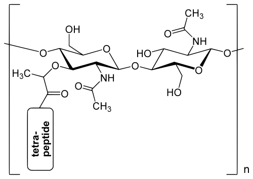
細菌の細胞壁内のペプチドグリカン層は、N-アセチルグルコサミン(GlcNAcまたはNAG)とN-アセチルムラミン酸(MurNAcまたはNAM)という2種類のアミノ糖が交互に並んだ直鎖から形成される結晶格子構造である。これらの糖はβ-(1,4)-グリコシド結合で連結されている。各MurNAcは、大腸菌(グラム陰性菌)の場合はL-アラニン、D-グルタミン酸、メソ-ジアミノピメリン酸、およびD-アラニンを含む短い(4~5残基)アミノ酸鎖に結合している。一方、黄色ブドウ球菌(グラム陽性菌)の場合は、L-アラニン、D-グルタミン、L-リジン、およびD-アラニンと、テトラペプチド間に5-グリシン架橋を持つアミノ酸鎖に結合している。ペプチドグリカンは、自然界におけるD-アミノ酸の最も重要な供給源の一つである。
ペプチドグリカン層は内膜を囲むことで、細胞の膨圧によって引き起こされる溶解から細胞を保護します。細胞壁が成長すると、その形状は生涯を通じて維持されるため、棒状の細胞は棒状の細胞のままであり、球状の細胞は生涯にわたって球状の細胞のままです。これは、新たに合成された隔壁物質が、子孫細胞のために半球状の壁に変化するためです。[ 9 ]
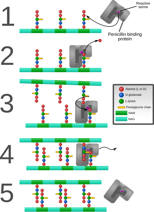
異なる直鎖アミノ糖鎖のアミノ酸間の架橋は、酵素DD-トランスペプチダーゼの助けを借りて起こり、強くて剛性のある3次元構造をもたらします。特定のアミノ酸配列と分子構造は、細菌種によって異なります。[ 10 ]
The different peptidoglycan types of bacterial cell walls and their taxonomic implications have been described.[11]Archaea (domainArchaea)[12] do not contain peptidoglycan (murein).[13] Some Archaea contain pseudopeptidoglycan (pseudomurein, see below).[14]
Peptidoglycan is involved in binary fission during bacterial cell reproduction. L-form bacteria and mycoplasmas, both lacking peptidoglycan cell walls, do not proliferate by binary fission, but by a budding mechanism.[15][16]
In the course of early evolution, the successive development of boundaries (membranes, walls) protecting first structures of life against their environment must have been essential for the formation of the first cells (cellularisation).
The invention of rigid peptidoglycan (murein) cell walls in bacteria (domain Bacteria[12]) was probably the prerequisite for their survival, extensive radiation and colonisation of virtually all habitats of the geosphere and hydrosphere.[17][18]
The peptidoglycan monomers are synthesized in the cytosol and are then attached to a membrane carrier bactoprenol. Bactoprenol transports peptidoglycan monomers across the cell membrane where they are inserted into the existing peptidoglycan.[19]
Each of these reactions requires the energy source ATP.[20] This is all referred to as Stage one.
Stage two occurs in the cytoplasmic membrane. It is in the membrane where a lipid carrier called bactoprenol carries peptidoglycan precursors through the cell membrane.
In some archaea, i.e. members of the Methanobacteriales and in the genus Methanopyrus, pseudopeptidoglycan (pseudomurein) has been found.[14] In pseudopeptidoglycan the sugar residues are β-(1,3) linked N-acetylglucosamine and N-acetyltalosaminuronic acid. This makes the cell walls of such archaea insensitive to lysozyme.[23] The biosynthesis of pseudopeptidoglycan has been described.[24]
Peptidoglycan recognition is an evolutionarily conserved process.[25] The overall structure is similar between bacterial species, but various modifications can increase the diversity. These include modifications of the length of sugar polymers, modifications in the sugar structures, variations in cross-linking or substitutions of amino acids (primarily at the third position).[25][26] The aim of these modifications is to alter the properties of the cell wall, which plays a vital role in pathogenesis.[25]
Peptidoglycans can be degraded by several enzymes (lysozyme, glucosaminidase, endopeptidase...[25]), producing immunostimulatory fragments (sometimes called muropeptides[27]) that are critical for mediating host-pathogen interactions.[26] These include muramyl dipeptide (MDP), N-acetylglucosamine (NAG), or γ-d-glutamyl-meso-diaminopimelic acid (iE-DAP).[25][27]
Peptidoglycan from intestinal bacteria (both pathogens and commensals) crosses the intestinal barrier even under physiological conditions.[27] Mechanisms through which peptidoglycan or its fragments enter the host cells can be direct (carrier-independent) or indirect (carrier-dependent), and they are either bacteria-mediated (secretion systems, membrane vesicles) or host cell-mediated (receptor-mediated, peptide transporters).[27]Bacterial secretion systems are protein complexes used for the delivery of virulence factors across the bacterial cell envelope to the exterior environment.[28] Intracellular bacterial pathogens invade eukaryotic cells (which may lead to the formation of phagolysosomes and/or autophagy activation), or bacteria may be engulfed by phagocytes (macrophages, monocytes, neutrophils...). The bacteria-containing phagosome may then fuse with endosomes and lysosomes, leading to degradation of bacteria and generation of polymeric peptidoglycan fragments and muropeptides.[27]
Innate immune system senses intact peptidoglycan and peptidoglycan fragments using numerous PRRs (pattern recognition receptors) that are secreted, expressed intracellularly or expressed on the cell surface.[25]
PGLYRPs are conserved from insects to mammals.[27] Mammals produce four secreted soluble peptidoglycan recognition proteins (PGLYRP-1, PGLYRP-2, PGLYRP-3 and PGLYRP-4) that recognize muramyl pentapeptide or tetrapeptide.[25] They can also bind to LPS and other molecules by using binding sites outside of the peptidoglycan-binding groove.[28] After recognition of peptidoglycan, PGLYRPs activate polyphenol oxidase (PPO) molecules, Toll, or immune deficiency (IMD) signalling pathways. That leads to production of antimicrobial peptides (AMPs).[28]
Each of the mammalian PGLYRPs display unique tissue expression patterns. PGLYRP-1 is mainly expressed in the granules of neutrophils and eosinophils.[25] PGLYRP-3 and 4 are expressed by several tissues such as skin, sweat glands, eyes or the intestinal tract.[27] PGLYRP-1, 3 and 4 form disulphide-linked homodimers and heterodimers essential for their bactericidal activity.[27] Their binding to bacterial cell wall peptidoglycans can induce bacterial cell death by interaction with various bacterial transcriptional regulatory proteins.[25] PGLYRPs are likely to assist in bacterial killing by cooperating with other PRRs to enhance recognition of bacteria by phagocytes.[25]
PGLYRP-2 is primarily expressed by the liver and secreted into the circulation.[25] Also, its expression can be induced in skin keratinocytes, oral and intestinal epithelial cells.[27] In contrast with the other PGLYRPs, PGLYRP-2 has no direct bactericidal activity. It possesses peptidoglycan amidase activity, it hydrolyses the lactyl-amide bond between the MurNAc and the first amino acid of the stem peptide of peptidoglycan.[25][27] It is proposed, that the function of PGLYRP-2 is to prevent over-activation of the immune system and inflammation-induced tissue damage in response to NOD2 ligands (see below), as these muropeptides can no longer be recognized by NOD2 upon separation of the peptide component from MurNAc.[27] Growing evidence suggests that peptidoglycan recognition protein family members play a dominant role in the tolerance of intestinal epithelial cells toward the commensal microbiota.[28][29] It has been demonstrated that expression of PGLYRP-2 and 4 can influence the composition of the intestinal microbiota.[28]
Recently, it has been discovered, that PGLYRPs (and also NOD-like receptors and peptidoglycan transporters) are highly expressed in the developing mouse brain.[30] PGLYRP-2 and is highly expressed in neurons of several brain regions including the prefrontal cortex, hippocampus, and cerebellum, thus indicating potential direct effects of peptidoglycan on neurons. PGLYRP-2 is highly expressed also in the cerebral cortex of young children, but not in most adult cortical tissues. PGLYRP-1 is also expressed in the brain and continues to be expressed into adulthood.[30]
Probably the most well-known receptors of peptidoglycan are the NOD-like receptors (NLRs), mainly NOD1 and NOD2. The NOD1 receptor is activated after iE-DAP (γ-d-glutamyl-meso-diaminopimelic acid) binding, while NOD2 recognizes MDP (muramyl dipeptide), by their LRR domains.[28] Activation leads to self-oligomerization, resulting in activation of two signalling cascades. One triggers activation of NF-κB (through RIP2, TAK1 and IKK[31]), second leads to MAPK signalling cascade. Activation of these pathways induces production of inflammatory cytokines and chemokines.[25]
NOD1 is expressed by diverse cell types, including myeloid phagocytes, epithelial cells[25] and neurons.[30] NOD2 is expressed in monocytes and macrophages, epithelial intestinal cells, Paneth cells, dendritic cells, osteoblasts, keratinocytes and other epithelial cell types.[27] As cytosolic sensors, NOD1 and NOD2 must either detect bacteria that enter the cytosol, or peptidoglycan must be degraded to generate fragments that must be transported into the cytosol for these sensors to function.[25]
Recently, it was demonstrated that NLRP3 is activated by peptidoglycan, through a mechanism that is independent of NOD1 and NOD2.[27] In macrophages, N-acetylglucosamine generated by peptidoglycan degradation was found to inhibit hexokinase activity and induce its release from the mitochondrialmembrane. It promotes NLRP3 inflammasome activation through a mechanism triggered by increased mitochondrial membrane permeability.[27]
NLRP1 is also considered as a cytoplasmic sensor of peptidoglycan. It can sense MDP and promote IL-1 secretion through binding NOD2.[28][26]
C-type lectins are a diverse superfamily of mainly Ca2+-dependent proteins that bind a variety of carbohydrates (including the glycan skeleton of peptidoglycan), and function as innate immune receptors.[27] CLR proteins that bind to peptidoglycan include mannose binding lectin (MBL), ficolins, Reg3A (regeneration gene family protein 3A), and PTCLec1.[28] In mammals, they initiate the lectin-pathway of the complement cascade.[27]
The role of toll-like receptors (TLRs) in direct recognition of peptidoglycan is controversial.[25] In some studies, has been reported that peptidoglycan is sensed by TLR2.[32] But this TLR2-inducing activity could be due to cell wall lipoproteins and lipoteichoic acids that commonly co-purify with peptidoglycan. Also variation in peptidoglycan structure in bacteria from species to species may contribute to the differing results on this topic.[25][27]
Peptidoglycan is immunologically active, which can stimulate immune cells to increase the expression of cytokines and enhance antibody-dependent specific response when combined with vaccine or as adjuvant alone.[28] MDP, which is the basic unit of peptidoglycan, was initially used as the active component of Freund's adjuvant.[28] Peptidoglycan from Staphylococcus aureus was used as a vaccine to protect mice, showing that after vaccine injection for 40 weeks, the mice survived from S. aureus challenge at an increased lethal dose.[33]
Some antibacterial drugs such as penicillin interfere with the production of peptidoglycan by binding to bacterial enzymes known as penicillin-binding proteins or DD-transpeptidases.[6] Penicillin-binding proteins form the bonds between oligopeptide crosslinks in peptidoglycan. For a bacterial cell to reproduce through binary fission, more than a million peptidoglycan subunits (NAM-NAG+oligopeptide) must be attached to existing subunits.[34] Mutations in genes coding for transpeptidases that lead to reduced interactions with an antibiotic are a significant source of emerging antibiotic resistance.[35] Since peptidoglycan is also lacking in L-form bacteria and in mycoplasmas, both are resistant against penicillin.
Other steps of peptidoglycan synthesis can also be targeted. The topical antibiotic bacitracin targets the utilization of C55-isoprenyl pyrophosphate. Lantibiotics, which include the food preservative nisin, attack lipid II.[36]
Lysozyme, which is found in tears and constitutes part of the body's innate immune system exerts its antibacterial effect by breaking the β-(1,4)-glycosidic bonds in peptidoglycan (see above). Lysozyme is more effective in acting against gram-positive bacteria, in which the peptidoglycan cell wall is exposed, than against gram-negative bacteria, which have an outer layer of LPS covering the peptidoglycan layer.[31] Several bacterial peptidoglycan modifications can result in resistance to degradation by lysozyme. Susceptibility of bacteria to degradation is also considerably affected by exposure to antibiotics. Exposed bacteria synthesize peptidoglycan that contains shorter sugar chains that are poorly crosslinked and this peptidoglycan is then more easily degraded by lysozyme.[28]
2025年の研究では、溶解した細菌細胞から放出されたペプチドグリカン断片が、多様な細菌種間で一般的な危険信号として機能することが報告されている。[ 37 ]外因性ペプチドグリカンへの曝露は、 Vibrio cholerae、Pseudomonas aeruginosa、Staphylococcus aureus、Acinetobacter baumannii、Enterococcus faecalisにおいて、三次元バイオフィルムの形成を急速に誘導することが示された。短時間の曝露でも、バイオフィルムマトリックス成分の産生増加につながる制御応答を引き起こすのに十分であった。V . choleraeでは、ペプチドグリカンへの曝露により、バイオフィルム構造に寄与するvps -Iおよびvps -II遺伝子クラスターを含む、マトリックス合成に関与するいくつかの遺伝子が上方制御された。細菌が細胞外ペプチドグリカン断片を感知するメカニズムは依然として不明である。[ 37 ]