
Loading article…

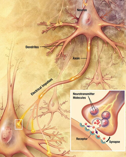
化学シナプスは、ニューロンの信号が互いに、また筋肉や腺などの非神経細胞に伝達される生物学的接合部です。化学シナプスによって、ニューロンは中枢神経系内で回路を形成することができます。これらは、知覚や思考の根底にある生物学的計算に不可欠です。また、神経系が体の他のシステムと接続し、制御することを可能にします。
化学シナプスでは、一方のニューロンがシナプス後細胞(例えば、別のニューロン)に隣接する小さな空間(シナプス間隙)に神経伝達物質分子を放出します。神経伝達物質分子はシナプス小胞と呼ばれる小さな袋の中に含まれており、エキソサイトーシスによってシナプス間隙に放出されます。これらの分子はシナプス後細胞上の神経伝達物質受容体に結合します。最後に、神経伝達物質は、酵素分解や、特定の輸送体によるシナプス前細胞または神経膠細胞への再取り込みなど、いくつかのメカニズムのいずれかによってシナプス間隙から除去され、その作用が終結します。
成人の人間の 脳には、10 14~ 5 × 10 14 (100 ~ 500 兆) 個のシナプスが含まれていると推定されています。[ 1 ]大脳皮質の 1 立方ミリメートルあたりには、およそ 10 億 (短いスケール、つまり 10 9 ) 個のシナプスが含まれています。[ 2 ]人間の大脳皮質のシナプスの数は、0.15 京 (150 兆) と別途推定されています。[ 3 ]
「シナプス」という言葉は、1897年にチャールズ・スコット・シェリントン卿によって導入されました。 [ 4 ]化学シナプスは生物学的シナプスの唯一の種類ではなく、電気シナプスや免疫シナプスも存在します。しかし、修飾語がない場合、「シナプス」は一般的に化学シナプスを指します。
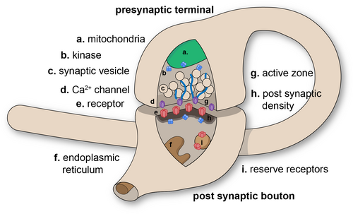
シナプスは、ニューロン間、またはニューロンと他の種類の細胞間の機能的な接続です。[ 5 ] [ 6 ] 典型的なニューロンは数千個のシナプスを形成しますが、はるかに少ないシナプスを形成するタイプもあります。[ 7 ] ほとんどのシナプスは軸索と樹状突起を接続しますが、[ 8 ] [ 9 ]軸索と細胞体、軸索同士、[ 10 ] [ 11 ]および樹状突起同士の接続など、他のタイプの接続もあります。[ 9 ]シナプスは、2 つの細胞の膜が接触しているように見える点を除いて、光学顕微鏡では認識するには小さすぎますが、電子顕微鏡を使用すると、その細胞要素を明確に視覚化できます。
化学シナプスは、シナプス前細胞からシナプス後細胞へ情報を一方向に伝達するため、構造と機能において非対称である。シナプス前軸索終末、またはシナプスシナプスボタンは、シナプス前細胞の軸索内の特殊な領域であり、シナプス小胞と呼ばれる小さな膜で囲まれた球状の構造物(およびミトコンドリアや小胞体などの他の多くの支持構造や細胞小器官)の中に神経伝達物質が含まれています。一部のシナプス小胞は、活動帯と呼ばれる領域のシナプス前細胞膜にドッキングしています。
そのすぐ反対側には、神経伝達物質受容体を含むシナプス後細胞領域があります。2つのニューロン間のシナプスでは、シナプス後領域は樹状突起または細胞体上に存在することがあります。シナプス後膜のすぐ後ろには、シナプス後密度(PSD)と呼ばれる、相互に連結したタンパク質の複雑な複合体があります。
PSDに含まれるタンパク質は、神経伝達物質受容体の固定と輸送、およびこれらの受容体の活性調節に関与している。受容体とPSDは、樹状突起の主幹から突き出た特殊な突起である樹状突起棘に多く見られる。
シナプスは対称型と非対称型に分類される。電子顕微鏡で観察すると、非対称型シナプスはシナプス前細胞に丸みを帯びた小胞を持ち、シナプス後部に目立つ密度構造が見られるのが特徴である。非対称型シナプスは一般的に興奮性である。一方、対称型シナプスは小胞が扁平または細長く、シナプス後部に目立つ密度構造は見られない。対称型シナプスは一般的に抑制性である。
シナプス間隙(シナプスギャップとも呼ばれる)は、シナプス前細胞とシナプス後細胞の間の隙間で、幅は約20 nm(0.02 μ)である。[ 12 ]間隙の体積が小さいため、神経伝達物質の濃度を迅速に上げ下げすることができる。[ 13 ]
オートシナプスとは、あるニューロンの軸索が自身の樹状突起とシナプスを形成する際に生じる化学的(または電気的)シナプスのことである。
一部の著者は、化学シナプスにおける信号伝達をパラクリンシグナル伝達の特殊なケースとみなしている[ 14 ]が、他の著者はそれを独立したシグナル伝達機構として扱っている[ 15 ] 。
以下に、シナプス前ニューロンからシナプス後細胞へのシナプス伝達で起こる一連の事象の概要を示します。各ステップについては、以下でさらに詳しく説明します。最終ステップを除き、最も速いシナプスでは、プロセス全体がわずか数百マイクロ秒で完了することに注意してください。[ 16 ]
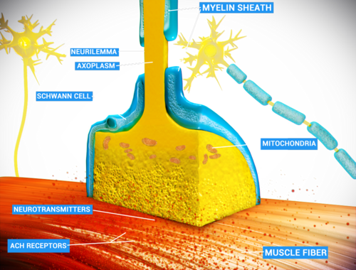
The release of a neurotransmitter is triggered by the arrival of a nerve impulse (or action potential) and occurs through an unusually rapid process of cellular secretion (exocytosis). Within the presynaptic nerve terminal, vesicles containing neurotransmitter are localized near the synaptic membrane. The arriving action potential produces an influx of calcium ions through voltage-dependent, calcium-selective ion channels at the down stroke of the action potential (tail current).[17] Calcium ions then bind to synaptotagmin proteins found within the membranes of the synaptic vesicles, allowing the vesicles to fuse with the presynaptic membrane.[18] The fusion of a vesicle is a stochastic process, leading to frequent failure of synaptic transmission at the very small synapses that are typical for the central nervous system. Large chemical synapses (e.g. the neuromuscular junction), on the other hand, have a synaptic release probability, in effect, of 1. Vesicle fusion is driven by the action of a set of proteins in the presynaptic terminal known as SNAREs. As a whole, the protein complex or structure that mediates the docking and fusion of presynaptic vesicles is called the active zone.[19] The membrane added by the fusion process is later retrieved by endocytosis and recycled for the formation of fresh neurotransmitter-filled vesicles.
An exception to the general trend of neurotransmitter release by vesicular fusion is found in the type II receptor cells of mammalian taste buds. Here the neurotransmitter ATP is released directly from the cytoplasm into the synaptic cleft via voltage gated channels.[20]
Receptors on the opposite side of the synaptic gap bind neurotransmitter molecules. Receptors can respond in either of two general ways. First, the receptors may directly open ligand-gated ion channels in the postsynaptic cell membrane, causing ions to enter or exit the cell and changing the local transmembrane potential.[16] The resulting change in voltage is called a postsynaptic potential. In general, the result is excitatory in the case of depolarizing currents, and inhibitory in the case of hyperpolarizing currents. Whether a synapse is excitatory or inhibitory depends on what type(s) of ion channel conduct the postsynaptic current(s), which in turn is a function of the type of receptors and neurotransmitter employed at the synapse. The second way a receptor can affect membrane potential is by modulating the production of chemical messengers inside the postsynaptic neuron. These second messengers can then amplify the inhibitory or excitatory response to neurotransmitters.[16]
After a neurotransmitter molecule binds to a receptor molecule, it must be removed to allow for the postsynaptic membrane to continue to relay subsequent EPSPs and/or IPSPs. This removal can happen through one or more processes:
The strength of a synapse has been defined by Bernard Katz as the product of (presynaptic) release probability pr, quantal size q (the postsynaptic response to the release of a single neurotransmitter vesicle, a 'quantum'), and n, the number of release sites. "Unitary connection" usually refers to an unknown number of individual synapses connecting a presynaptic neuron to a postsynaptic neuron. The amplitude of postsynaptic potentials (PSPs) can be as low as 0.4 mV to as high as 20 mV.[22] The amplitude of a PSP can be modulated by neuromodulators or can change as a result of previous activity. Changes in the synaptic strength can be short-term, lasting seconds to minutes, or long-term (long-term potentiation, or LTP), lasting hours. Learning and memory are believed to result from long-term changes in synaptic strength, via a mechanism known as synaptic plasticity.
Desensitization of the postsynaptic receptors is a decrease in response to the same neurotransmitter stimulus. It means that the strength of a synapse may in effect diminish as a train of action potentials arrive in rapid succession – a phenomenon that gives rise to the so-called frequency dependence of synapses. The nervous system exploits this property for computational purposes, and can tune its synapses through such means as phosphorylation of the proteins involved.
Synaptic transmission can be changed by previous activity. These changes are called synaptic plasticity and may result in either a decrease in the efficacy of the synapse, called depression, or an increase in efficacy, called potentiation. These changes can either be long-term or short-term. Forms of short-term plasticity include synaptic fatigue or depression and synaptic augmentation. Forms of long-term plasticity include long-term depression and long-term potentiation. Synaptic plasticity can be either homosynaptic (occurring at a single synapse) or heterosynaptic (occurring at multiple synapses).
Homosynaptic plasticity (or also homotropic modulation) is a change in the synaptic strength that results from the history of activity at a particular synapse. This can result from changes in presynaptic calcium as well as feedback onto presynaptic receptors, i.e. a form of autocrine signaling. Homosynaptic plasticity can affect the number and replenishment rate of vesicles or it can affect the relationship between calcium and vesicle release. Homosynaptic plasticity can also be postsynaptic in nature. It can result in either an increase or decrease in synaptic strength.
One example is neurons of the sympathetic nervous system (SNS), which release noradrenaline, which, besides affecting postsynaptic receptors, also affects presynaptic α2-adrenergic receptors, inhibiting further release of noradrenaline.[23] This effect is utilized with clonidine to perform inhibitory effects on the SNS.
Heterosynaptic plasticity (or also heterotropic modulation) is a change in synaptic strength that results from the activity of other neurons. Again, the plasticity can alter the number of vesicles or their replenishment rate or the relationship between calcium and vesicle release. Additionally, it could directly affect calcium influx. Heterosynaptic plasticity can also be postsynaptic in nature, affecting receptor sensitivity.
One example is again neurons of the sympathetic nervous system, which release noradrenaline, which, in addition, generates an inhibitory effect on presynaptic terminals of neurons of the parasympathetic nervous system.[23]
In general, if an excitatory synapse is strong enough, an action potential in the presynaptic neuron will trigger an action potential in the postsynaptic cell. In many cases the excitatory postsynaptic potential (EPSP) will not reach the threshold for eliciting an action potential. When action potentials from multiple presynaptic neurons fire simultaneously, or if a single presynaptic neuron fires at a high enough frequency, the EPSPs can overlap and summate. If enough EPSPs overlap, the summated EPSP can reach the threshold for initiating an action potential. This process is known as summation, and can serve as a high pass filter for neurons.[24]
一方、GABAなどの抑制性神経伝達物質を放出するシナプス前ニューロンは、シナプス後ニューロンに抑制性シナプス後電位(IPSP)を引き起こし、膜電位を閾値から遠ざけ、興奮性を低下させ、ニューロンが活動電位を発生させることをより困難にする。IPSPがEPSPと重なる場合、多くの場合、IPSPはニューロンが活動電位を発火するのを阻止することができる。このように、ニューロンの出力は、多くの異なるニューロンからの入力に依存する可能性があり、それぞれのニューロンは、そのニューロンとのシナプスの強度と種類に応じて、異なる程度の影響を及ぼす可能性がある。ジョン・カリュー・エクルズは、シナプス統合に関する重要な初期の実験のいくつかを行い、 1963年にノーベル生理学・医学賞を受賞した。
神経伝達物質がシナプスで放出されると、シナプス間隙の狭い空間内で最高濃度に達しますが、再吸収または分解される前に一部は拡散して失われます。拡散した場合、他のシナプスまたはシナプスから離れた膜上に存在する受容体を活性化する可能性があります。神経伝達物質のシナプス外活動はボリューム伝達として知られています。[ 25 ] このような効果がある程度発生することはよく知られていますが、その機能的重要性は長い間議論の的となってきました。[ 26 ]
最近の研究では、ボリューム伝達が一部の特殊なタイプのニューロンの主要な相互作用様式である可能性が示唆されている。哺乳類の脳皮質では、神経膠様細胞と呼ばれるニューロンのクラスが、神経伝達物質GABAを細胞外空間に放出することで、近くの他の皮質ニューロンを抑制することができる。[ 27 ]神経膠様細胞から細胞外空間に放出されたGABAは、周囲のアストロサイトにも作用し、ボリューム伝達がイオンと神経伝達物質の恒常性の制御において役割を果たしていることを示している。[ 28 ]神経膠様細胞のボタンの約78%は、古典的なシナプスを形成しない。これは、古典的なシナプスが存在しない場所でニューロンが化学的に通信する最初の明確な例かもしれない。[ 27 ]
An electrical synapse is an electrically conductive link between two abutting neurons that is formed at a narrow gap between the pre- and postsynaptic cells, known as a gap junction. At gap junctions, cells approach within about 3.5 nm of each other, rather than the 20 to 40 nm distance that separates cells at chemical synapses.[29][30] As opposed to chemical synapses, the postsynaptic potential in electrical synapses is not caused by the opening of ion channels by chemical transmitters, but rather by direct electrical coupling between both neurons. Electrical synapses are faster than chemical synapses.[13] Electrical synapses are found throughout the nervous system, including in the retina, the reticular nucleus of the thalamus, the neocortex, and in the hippocampus.[31] While chemical synapses are found between both excitatory and inhibitory neurons, electrical synapses are most commonly found between smaller local inhibitory neurons. Electrical synapses can exist between two axons, two dendrites, or between an axon and a dendrite.[32][33] In some fish and amphibians, electrical synapses can be found within the same terminal of a chemical synapse, as in Mauthner cells.[34]
化学シナプスの最も重要な特徴の1つは、ほとんどの精神活性薬の作用部位であることです。シナプスは、クラーレ、ストリキニーネ、コカイン、モルヒネ、アルコール、LSD、リスペリドンなど、数え切れないほどの薬物の影響を受けます。これらの薬物はシナプス機能に異なる影響を与え、多くの場合、特定の神経伝達物質を使用するシナプスに限定されます。たとえば、クラーレはアセチルコリンがシナプス後膜を脱分極するのを阻害し、麻痺を引き起こす毒物です。ストリキニーネは神経伝達物質グリシンの抑制効果を阻害し、体がこれまで無視していた弱い刺激に反応するようになり、制御不能な筋肉の痙攣を引き起こします。モルヒネはエンドルフィン神経伝達物質を使用するシナプスに作用し、アルコールは神経伝達物質GABAの抑制効果を高めます。LSDは神経伝達物質セロトニンを利用するシナプスに干渉します。リスペリドンは、ドーパミン受容体やセロトニン受容体など、さまざまな受容体の遮断薬であり、一部のセロトニン受容体には高い親和性で結合します。コカインはドーパミンの再取り込みを阻害するため、その効果を増強します。
1950年代、バーナード・カッツとポール・ファットはカエルの神経筋接合部で自発的な微小シナプス電流を観察した。[ 35 ]これらの観察に基づいて、彼らは「量子仮説」を提唱し、これが神経伝達物質放出をエキソサイトーシスとして理解する現在の基礎となり、カッツはこの功績により1970年にノーベル生理学・医学賞を受賞した。[ 36 ] 1960年代後半、リカルド・ミレディとカッツは、脱分極によって誘発されるカルシウムイオンの流入がエキソサイトーシスを引き起こすという仮説を提唱した。
チャールズ・スコット・シェリントン卿は「シナプス」という言葉を造語し、その語源については、シェリントンがジョン・フルトンに宛てた手紙の中で述べている。
「神経細胞と神経細胞の接合部を呼ぶ名前が必要だと感じました...私は「シンデスム」を使うことを提案しました...彼[サー・マイケル・フォスター]は、エウリピデス研究者であるトリニティ大学の友人ヴェラルに相談し、ヴェラルは「シナプス」(ギリシャ語の「留め金」から)を提案しました。」–チャールズ・スコット・シェリントン[ 4 ]
synapses connect axons to cell body.
{{cite book}}: CS1 maint: url-status (link)