
Loading article…
Dry eye disease (DED), also known as keratoconjunctivitis sicca, is the condition of having dryeyes.[2] The term dry eye syndrome was formerly used, but is now avoided following advances that have established it as a distinct disease entity.[6]
Symptoms of dry eye include dryness in the eye, irritation, redness, discharge, blurred vision, and easily fatigued eyes. Symptoms range from mild and occasional to severe and continuous.[3] DED can lead to blurred vision, instability of the tear film, increased risk of damage to the ocular surface such as scarring of the cornea, and changes in the eye including the neurosensory system.[2][7]
Dry eye occurs either when the eye does not produce enough tears or when the tears evaporate too quickly.[2] This can be caused by age, contact lens use, meibomian gland dysfunction,[8]pregnancy, Sjögren's disease, vitamin A deficiency, omega-3 fatty acid deficiency, LASIK surgery, and certain medications such as antihistamines, some blood pressure medication, hormone replacement therapy, and antidepressants.[2][3][4] Chronic conjunctivitis such as from tobacco smoke exposure or infection may also lead to the condition.[2] Diagnosis is mostly based on the symptoms, though several other tests may be used.[9] Dry eye disease occasionally makes wearing contact lenses impossible.[2]
治療は根本原因によって異なります。通常、人工涙液が第一選択薬です。顔にぴったりフィットするラップアラウンド型の眼鏡は涙の蒸発を減らす可能性があります。[ 10 ]服用している薬を注意深く調べ、安全であれば薬を変更することで、これらの薬が原因である場合は症状が改善することもあります。局所薬、つまり点眼薬が症状の治療に役立つ場合があります。免疫抑制剤のシクロスポリン(シクロスポリン)は涙の分泌を増やすために推奨される場合があり、短期使用の場合は局所コルチコステロイド薬も炎症を軽減するのに役立つことがあります。[ 7 ]
ドライアイは一般的な眼疾患です。[ 3 ]調査対象集団によって異なりますが、5~34%の人が何らかの影響を受けています。[ 5 ]高齢者では最大70%が影響を受けています。[ 11 ]中国では約17%の人が影響を受けています。[ 12 ] 「keratoconjunctivitis sicca」という語は、ラテン語で「角膜と結膜の乾燥」を意味します。[ 13 ]
涙液膜・眼表面学会ドライアイワークショップ(TFOS DEWS)II報告書(2017年)[ 14 ]では、ドライアイは多分野にわたる国際的な委員会によって次のように定義されました。
TFOS DEWS III (2025) によると、[ 6 ]疾患病理学と涙液膜に関する最新の理解を踏まえると、定義を根本的に変更する必要はないという点で合意が得られた。しかし、改訂された定義では、恒常性を維持する上で眼表面組織と涙液膜の両方が果たす本質的な役割が強調され、以下の更新された文言となった。
The TFOS DEWS definitions have contributed to establishing a clear characterization of dry eye as a disease. Whereas it was formerly described as a syndrome because of the limited understanding of its etiology, subsequent advances in research have clarified its diagnostic features, natural history, and therapeutic responses, supporting its recognition as a distinct disease entity.[6]
Typical symptoms of dry eye disease are dryness,[15] burning[16] and a sandy-gritty eye irritation that gets worse as the day goes on.[17] Symptoms may also be described as itchy, stinging or tired eyes.[16][18] Other symptoms are pain, redness, a pulling sensation, and pressure behind the eye.[4][16] There may be a feeling that something, such as a speck of dirt, is in the eye.[4][16] The resultant damage to the eye's surface increases discomfort and sensitivity to bright light.[16] Both eyes usually are affected.[19]
There may also be a stringy discharge from the eyes. Although it may seem contradictory, dry eye can cause the eyes to water due to irritation. One may experience excessive tearing such as if something got into the eye. These reflex tears will not necessarily make the eyes feel better since they are the watery tears that are produced in response to injury, irritation, or emotion which lack the lubricating qualities necessary to prevent dry eye.[4]
まばたきは涙で目を覆うため、長時間目を使うことでまばたきの頻度が減る活動によって症状が悪化します。[ 16 ]これらの活動には、長時間の読書、コンピューターの使用(コンピューター視覚症候群)、運転、テレビの視聴などが含まれます。[ 4 ] [ 16 ]風が強い場所、ほこりっぽい場所、煙(タバコの煙を含む)の多い場所、飛行機などの高高度の乾燥した環境、湿度の低い日、エアコン(特に車内)、扇風機、ヒーター、またはヘアドライヤーを使用している場所では症状が悪化します。[ 4 ] [ 16 ] [ 17 ] [ 19 ]涼しい、雨の降る、霧の降る天候や、シャワーなどの湿度の高い場所では症状が軽減します。[ 16 ]
ドライアイのほとんどの人は、長期的な影響のない軽度の刺激を経験します。しかし、この状態が治療されずに放置されたり、重症化すると、眼の損傷、涙液膜の不安定性、神経感覚の変化、視力障害、または(まれに)視力喪失を引き起こす合併症が生じる可能性があります。[ 4 ] [ 7 ]
コンピューター、スマートフォン、タブレット、その他のデジタル機器の画面を見すぎると、ドライアイを引き起こす可能性があります。[ 20 ]米国眼科学会は、「人間は通常、1分間に約15回まばたきをします。しかし、研究によると、コンピューターやその他のデジタル画面機器を使用している間は、1分間に約5〜7回しかまばたきをしません。まばたきは、目の表面に必要な水分を得るための目の動きです」と述べています。[ 20 ]
加齢は、涙の分泌量が加齢とともに減少するため、ドライアイの最も一般的な原因の 1 つです。[ 4 ]処方薬と市販薬 (OTC) の両方のいくつかの種類の薬が、特に高齢者のドライアイの主な原因の 1 つであると推測されています。特に、口渇も引き起こす抗コリン薬は、ドライアイを促進すると考えられています。[ 21 ]ドライアイは、熱傷や化学熱傷、または (流行の場合)アデノウイルスによっても引き起こされる可能性があります。多くの研究で、糖尿病患者はドライアイのリスクが高いことがわかりました。[ 22 ]
About half of all people who wear contact lenses complain of dry eyes.[4] There are two potential connections between contact usage and dry eye. Traditionally, it was believed that soft contact lenses, which float on the tear film that covers the cornea, absorb the tears in the eyes.[4] The connection between a loss in nerve sensitivity and tear production is also the subject of current research.[23]
Dry eye also occurs or becomes worse after laser-assisted in situ keratomileusis (LASIK) and other refractive surgeries, in which the corneal nerves that stimulate tear secretion[4] are cut during the creation of a corneal flap.[4] Dry eye resulting from these procedures usually resolves after several months, but it can be permanent.[19] Individuals considering refractive surgery should be aware of this potential complication.[4]
An eye injury or other problem with the eyes or eyelids, such as bulging eyes or a drooping eyelid may lead to keratoconjunctivitis sicca.[18] Eyelid disorders may disrupt the complex blinking motion necessary to distribute tears evenly.[19]
Abnormalities of the mucin tear layer resulting from vitamin A deficiency, trachoma, diphtheric keratoconjunctivitis, mucocutaneous disorders, and certain topical medications are also implicated in keratoconjunctivitis sicca.[17]
Individuals with keratoconjunctivitis sicca exhibit elevated levels of tear nerve growth factor (NGF).[17] NGF on the ocular surface may play a significant role in the inflammation associated with dry eyes.[17]
Seasonal variations in the manifestation of dry eye have also been reported.[24]
The use of eye make-up products is another influencing factor. Although eye cosmetics have a long history and have been investigated for years, comprehensive reviews on their role in dry eye disease[25] and in ocular surface and adnexal disease[26] were first published in 2022 and 2023, respectively.
ドライアイがしばらく続くと、眼の表面に小さな擦過傷が生じることがあります。 [ 18 ]進行した症例では、上皮に病理学的変化、すなわち扁平上皮化生と杯細胞の消失が起こります。[ 17 ]重症例では、角膜表面の肥厚、角膜びらん、点状角膜症、上皮欠損、角膜潰瘍(無菌性および感染性)、角膜新生血管形成、角膜瘢痕、角膜菲薄化、さらには角膜穿孔に至ることもあります。[ 16 ] [ 17 ]
もう一つの要因として、ラクリチンモノマー欠乏が考えられます。ラクリチンの活性型であるラクリチンモノマーは、涙液欠乏性ドライアイ、シェーグレン症候群ドライアイ、コンタクトレンズ関連ドライアイ、および眼瞼炎で選択的に減少します。[ 27 ]多様な微生物群集で構成される眼表面マイクロバイオームは、ドライアイ疾患の病因に関与しており、眼表面の炎症や恒常性に影響を与える可能性があります。[ 28 ]
ドライアイの診断において、症状評価は重要な要素であり、ドライアイは症状に基づく疾患であると多くの人が考えているほどです。診断を可能にするスコアを決定するために、いくつかの質問票が開発されています。眼表面疾患指数(OSDI)は、臨床診療と研究において最も頻繁に使用される質問票です。[ 15 ]
一部の検査では、患者を「水分欠乏型」または「過剰蒸発型」の2つのカテゴリーのいずれかに分類することができます。診断ガイドラインは、2007年にドライアイワークショップ[ 5 ]によって発表され、 2017年にドライアイワークショップIIによって更新されました。[ 29 ] [ 30 ]スリットランプ検査は、ドライアイを診断し、眼の損傷を記録するために実施できます。[ 16 ] [ 17 ]この検査を実施する際、医師はまぶたの縁を検査します。[ 5 ]
シルマーテストは、眼を潤す水分量を測定できます。[ 16 ]このテストは、症状の重症度を判断するのに役立ちます。[ 4 ] 幅5mm、長さ35mmのワットマン#41ろ紙を使用して、麻酔ありとなしの両方で5分間のシルマーテストを 実施します。このテストでは、麻酔の有無にかかわらず、5mm未満の濡れが ドライアイの診断とみなされます。[ 17 ]
シルマーテストの結果が異常な場合は、反射性分泌を測定するためにシルマーIIテストを実施できます。このテストでは、綿棒で鼻粘膜を刺激した後、ワットマン#41ろ紙で涙の分泌量を測定します。このテストでは、 5分後に15mm未満の濡れが異常とみなされます。[ 17 ]
涙液破壊時間(TBUT)検査は、眼内の涙液が破壊されるまでの時間を測定するものです。[ 4 ]涙液破壊時間は、結膜嚢にフルオレセインを1滴滴下した後に測定できます。 [ 17 ] [ 5 ]
涙液タンパク質分析検査では、涙液中に含まれるリゾチームを測定します。涙液中のリゾチームは、総タンパク質含有量の約20~40パーセントを占めています。[ 17 ]
ラクトフェリン分析検査は他の検査と良好な相関関係を示す。[ 17 ]
最近発見された涙液中に自然に存在する分子Ap4Aは、さまざまな眼の乾燥状態において異常に高い濃度を示します。この分子は、単純なシルマーテストで涙液サンプルを採取するだけで生化学的に定量できます。この技術を利用することで、患者の涙液中のAp4A濃度を測定し、サンプルがドライアイを示しているかどうかを客観的に診断することが可能です。[ 31 ]
涙液浸透圧検査は、ドライアイ疾患の検査として提案されている。[ 32 ]涙液浸透圧は、角膜および結膜染色、涙液破壊時間、シルマーテスト、マイボーム腺グレードと比較して、ドライアイの診断および重症度分類においてより感度の高い方法である可能性がある。[ 33 ]一方で、ドライアイ治療のモニタリングにおける涙液浸透圧の有用性に疑問を呈する声もある。[ 27 ]
Any abnormality of any one of the three layers of tears produces an unstable tear film, resulting in symptoms of dry eyes.[17] Dry eye can be classified in two ways. The Tear Film & Ocular Surface Society Dry Eye Workshop (TFOS DEWS) II report offers a clinically relevant classification, while the Madrid triple classification is based on etiology, anatomical pathology, and clinical severity.[34][35] According to the TFOS DEWS II report, dry eye is broadly classified into two major types: (1) Aqueous-deficient dry eye (ADDE), which involves impaired lacrimal secretion, and (2) Evaporative dry eye (EDE), characterized by excessive tear loss from the ocular surface. However, many present with mixed forms of dry eye.[36][37]
The most common cause of dry eye is increased evaporation of the tear film (evaporative dry eye; EDE), typically as a result of meibomian gland dysfunction (MGD). The meibomian glands are two sets of oil glands that line the upper and lower eyelids and secrete the oily outer layer of the tear film—the lipid layer (TFLL). These glands often become clogged due to inflammation caused by blepharitis and/or rosacea, preventing an even distribution of oil (meibum). The result is an unstable lipid layer that is believed to increase evaporation of the tear film.[38] While the anti-evaporative function of the tear film as a whole is well established,[39] scientific evidence specifically supporting the TFLL as the primary source of this resistance remains mixed.[39][40][41] Another TFLL-associated mechanism is oxidative stress generated in the perturbed lipid layer due to altered meibum, based on a newly proposed function of the TFLL in corneal oxygenation by Mazyar Yazdani from Oslo University Hospital.[42]
ドライアイ(涙腺分泌低下症)による涙液産生不足が原因でドライアイ(涙液欠乏性ドライアイ;ADDE)が起こることがあります。[ 16 ] [ 17 ]涙腺が、結膜と角膜全体を完全な層で覆うのに十分な涙液を産生しないのです。[ 16 ]これは通常、他に健康上の問題がない人に起こります。加齢に伴い涙液分泌量が減少します。[ 17 ]これは閉経後の女性に最も多く見られるタイプです。[ 16 ] [ 43 ]
In many cases, aqueous deficient dry eye may have no apparent cause (idiopathic). Other causes include congenital alacrima, xerophthalmia, lacrimal gland ablation, and sensory denervation.[17] In rare cases, it may be a symptom of collagen vascular diseases, including relapsing polychondritis, rheumatoid arthritis, granulomatosis with polyangiitis, and systemic lupus erythematosus.[16][17][44][45]Sjögren syndrome and other autoimmune diseases are associated with aqueous tear deficiency.[16][17] Drugs such as isotretinoin, sedatives, diuretics, tricyclic antidepressants, antihypertensives, oral contraceptives, antihistamines, nasal decongestants, beta-blockers, phenothiazines, atropine, and pain relieving opiates such as morphine can cause or worsen this condition.[4][16][17] Infiltration of the lacrimal glands by sarcoidosis or tumors, or post-radiation fibrosis of the lacrimal glands can also cause this condition.[17] Recent attention has been paid to the composition of tears in normal or dry-eye individuals. Only a small fraction of the estimated 1543 proteins in tears are differentially deficient or upregulated in dry eye, one of which is lacritin.[46][27] Topical lacritin promotes tearing in rabbit preclinical studies.[47] Also, topical treatment of eyes of dry eye mice (Aire knockout mouse model of dry eye) restored tearing, and suppressed both corneal staining and the size of inflammatory foci in lacrimal glands.[48]
Avoiding refractive surgery (LASIK and PRK), limiting contact lens use, limiting computer screen use, and avoiding environmental conditions can decrease symptoms.[49] Complications can be prevented by use of wetting and lubricating drops and ointments.[50]
A variety of approaches can be taken to treat dry eye disease. Approaches include: avoidance of exacerbating factors (things that make it worse), tear stimulation and supplementation, increasing tear retention, eyelid cleansing, and treatment of eye inflammation.[51]
Conditions such as blepharitis can often co-exist and paying particular attention to cleaning the eyelids morning and night with mild soaps and warm compresses can improve both conditions.[51]
Dry eyes can be worsened by smoky environments, dust, and indoor air conditioning, and by our natural tendency to reduce our blink rate when concentrating. Purposefully blinking, especially during computer use, and resting tired eyes are basic steps that can be taken to minimise discomfort.[51] Rubbing one's eyes can irritate them further, so should be avoided.[19] Dry, drafty environments and those with smoke and dust should be avoided.[16] This includes avoiding hair dryers, heaters, air conditioners, or fans, especially when these devices are directed toward the eyes. Wearing glasses or directing gaze downward, for example, by lowering computer screens can be helpful to protect the eyes when aggravating environmental factors cannot be avoided.[19] Using a humidifier, especially in the winter, can help by adding moisture to the dry indoor air.[16][18][19][51]
For mild and moderate cases, supplemental lubrication is the most important part of treatment.[17] Application of artificial tears is sometimes suggested every few hours and may provide temporary relief.[16] Most artificial tear fluids contain mucoadhesive polymers such as hyaluronic acid, cellulose derivatives or polyvinyl alcohol as lubricants.[52] These polymers remain for a prolonged period of time on the ocular surface binding high amounts of water. By the covalent attachment of thiol groups to such polymers, their ocular residence time can be even improved, as thiolated polymers (thiomers) form disulfide bonds with cysteine-rich subdomains of mucus glycoproteins on the ocular surface.[53] Chitosan-N-acetylcysteine containing eye drops showed a significant reduction in symptoms of dry eye disease.[54] There are many different types of artificial tear on the market, however, there is no strong evidence to suggest that certain artificial tear formulations are superior to others in treating dry eye.[55]
Eye drops that include autologous serum (serum taken from the same person's blood and used in an eye drop formulation) are sometimes suggested to help supplement natural tears. The composition of serum has similarities to natural tears and may mimic natural tears. Evidence supporting this approach shows that autologous serum may be superior to artificial tears at relieving symptoms in the short-term, however, there is no strong evidence that autologous serum eye drops are better than artificial tears or saline solution for long-term symptom relief.[56]
潤滑性涙軟膏は日中にも使用できますが、塗布後に視界が悪くなるため、一般的には就寝時に使用されます。[ 17 ]白色ワセリン、鉱物油、および同様の潤滑剤が含まれています。 [ 17 ]潤滑剤および軟化剤として機能します。[ 17 ]塗布するには、下まぶたを下に引っ張り、少量(0.25 インチ)を内側に塗布する必要があります。[ 17 ]症状の重症度に応じて、1 時間ごとに塗布する場合もあれば、就寝時のみに塗布する場合もあります。[ 17 ]コンタクトレンズを装着している場合は絶対に使用しないでください。[ 17 ]目の周りに水分チャンバーを形成するように特別に設計された眼鏡を使用して、追加の湿度を作り出すことができます。[ 19 ]
涙液膜の浸透圧上昇に反応して起こる炎症は、軽度の局所コルチコステロイドまたはシクロスポリン(レスタシス、ヴェヴィエ)などの局所免疫抑制剤で抑制できます。 [ 7 ] [ 57 ] [ 58 ] [ 59 ] [ 60 ]涙液中のNGF濃度の上昇は、0.1%プレドニゾロンで低下させることができます。[ 17 ]
局所コルチコステロイドは、ドライアイの症状が炎症によって引き起こされている可能性がある人によく処方され、潤滑剤や人工涙液単独の治療と比較して、ドライアイの症状をわずかにまたは中程度改善する可能性があります。[ 7 ]局所コルチコステロイド治療が涙液膜の質や自然な涙の量を改善するかどうかは明らかではありません。[ 7 ]また、局所コルチコステロイド治療の長期使用には、眼圧上昇のリスク増加、白内障発症のリスク増加、眼感染症のリスク増加など、考慮すべきリスクもあります。ドライアイの局所コルチコステロイド治療から恩恵を受ける可能性のある人にとって、理想的な治療レジメン、局所製剤の処方、およびこの薬剤の潜在的なリスクのバランスは明らかではありません。[ 7 ]
Topical ciclosporin (topical ciclosporin A, tCSA) 0.05% ophthalmic emulsion is an immunosuppressant that is commonly used to treat symptoms of dry eye disease.[17][61] The drug decreases surface inflammation to increase tear production.[19] Some people find relief and report increased tear production, however, evidence of effectiveness from clinical trials is not strong and although some people may find relief, effectiveness may be inconsistent in different people.[61] Ciclosporin A treatment also comes with risks of adverse effects that are generally not serious but include a burning sensation.[61] Ciclosporin should not be used while wearing contact lenses,[17] during eye infections[4] or in people with a history of herpes virus infections.[19] Side effects include burning sensation (common),[4] redness, discharge, watery eyes, eye pain, foreign body sensation, itching, stinging, and blurred vision.[17][4] Long-term use of ciclosporin at high doses is associated with an increased risk of cancer.[62][63] Cheaper generic alternatives are available in some countries.[64]
天然の涙と人工の涙の両方をより長く持続させる方法がある。[ 19 ]
各眼には、涙を涙管に排出する小さな開口部である涙点[ 73 ]が2つあります[ 4 ] 。涙管を部分的または完全に閉じる方法があります[ 19 ] 。これにより、涙が鼻に流れ込むのを防ぎ、より多くの涙が目に供給されます[ 16 ] 。各眼の片方または両方の涙点への排出を遮断することができます。
涙点プラグは涙の排出を遮断するために涙点に挿入されます。[ 4 ]涙点プラグがドライアイの症状を軽減するのに効果的かどうかは明らかではありません。[ 74 ]涙点プラグは「比較的安全」と考えられていますが、その使用により流涙症(涙目)や、まれに涙が排出される涙嚢の重篤な感染症や腫れを引き起こす可能性があります。[ 74 ]これらは、他の医学的治療が不十分な中等度または重度のドライアイの患者に限定されています。[ 4 ]
涙点プラグが有効な場合は、涙点の熱焼灼[ 19 ]または電気焼灼[ 17 ] を行うことができる。熱焼灼では、局所麻酔薬を使用し、その後熱したワイヤーを適用する[ 19 ]。これにより、涙道の組織が収縮して瘢痕が形成され、涙管が閉じる[ 19 ]。
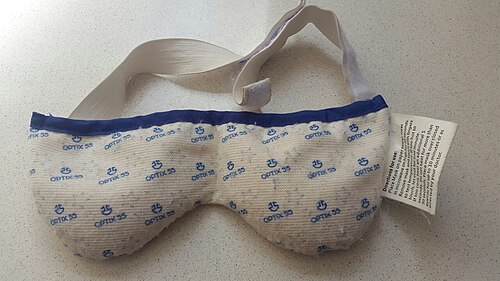
長鎖オメガ3サプリメントが役立つ可能性があるという証拠があるが[ 75 ]、プロバイオティクス、魚油、亜麻仁油、麻の実油(オメガ3)サプリメントは症状の緩和に効果がないようだ[ 76 ] [ 28 ] 。
BlephExは、眼瞼炎やドライアイ疾患に使用される医療機器です。 [ 77 ]この携帯型機器は、医師がまつ毛の生え際でまぶたの角質を除去し[ 78 ] 、慢性的なまぶたの疾患や不快感の原因となる炎症性バイオフィルム[ 79 ]を除去するために使用します。 [ 80 ]
For MGD, intense pulsed light (IPL) is a therapeutic modality that was originally developed for dermatological applications and later adopted in ophthalmology. IPL treatment has been shown to improve tear film stability, enhance meibomian gland function, and alleviate symptoms of ocular dryness.[81] Based on the 2020 comprehensive review of IPL,[81] the procedure shows considerable potential among reported studies in the literature. In 2021 in the United States, the Food and Drug Administration granted de novo authorization in the United States for an IPL device to manage dry eye disease due to meibomian gland dysfunction.[82]
In severe cases of dry eyes, tarsorrhaphy may be performed where the eyelids are partially sewn together. This reduces the palpebral fissure (eyelid separation), ideally leading to a reduction in tear evaporation.[16]
Keratoconjunctivitis sicca usually is a chronic problem.[19] Its prognosis shows considerable variance, depending upon the severity of the condition. Most people have mild-to-moderate cases, and can be treated symptomatically with lubricants. This provides an adequate relief of symptoms.[17]
When dry eye symptoms are severe, they can interfere with quality of life.[4] People sometimes feel their vision blurs with use, or severe irritation to the point that they have trouble keeping their eyes open or they may not be able to work or drive.[16][4]
Keratoconjunctivitis sicca is relatively common within the United States, especially in patients[17] aged 40 or older.[19] 10–20% of adults experience Keratoconjunctivitis sicca.[74] Approximately 1 to 4 million adults (age 65–84) in the US are affected.[74]
While persons with autoimmune diseases have a high likelihood of having dry eyes, most persons with dry eyes do not have an autoimmune disease.[19] Instances of Sjögren syndrome and keratoconjunctivitis sicca associated with it are present much more commonly in women, with a ratio of 9:1. In addition, milder forms of keratoconjunctivitis sicca also are more common in women.[17] This is partly because hormonal changes, such as those that occur in pregnancy, menstruation, and menopause, can decrease tear production.[4][19]
In areas of the world where malnutrition is common, vitamin A deficiency is a common cause. This is rare in the United States.[50]
Racial predilections do not exist for this disease.[17]
A study based on 274 answers using the Ocular Surface Disease Index (OSDI) from medical school students aged between 20 and 25 years old found a prevalence of dry eye symptoms of 83.6%.[83]
The following summarizes DED and MGD prevalence from TFOS DEWS III (2025),[84] highlighting findings from population-based studies and meta-analyses across different diagnostic approaches, stratified by age and sex.
Based on the Women's Health Study criteria, DED prevalence increases with age, from about 2.7% at 20–29 years to 30% in women over 80. The rate rises notably after 40 in both sexes, with women showing higher prevalence beyond 50. One study also reported high rates among individuals aged 10–19, without sex differences.
Based on signs and symptoms, DED prevalence ranges from 4.7% in children aged 6–9 to 62.9% in women aged 20–29, although confidence intervals are wide. Rates remain relatively consistent across adult age groups, with a decline in symptoms after 70. Sex differences are small except in those ≥70, where women show higher prevalence.
Based on TFOS DEWS II criteria, DED prevalence ranges from 5.4% in children aged 6–9 to 44.2% overall. Rates are comparable to those based on signs and symptoms. Above 30 years, prevalence is higher in females, while males show a clearer age-related increase.
Based on claims data, DED prevalence ranges from 2.8% to 8.5%, generally lower than estimates from clinical criteria. These figures derive from diagnostic or treatment codes in insurance or ICD data. Insufficient information was available to assess age- or sex-specific trends.
Based on clinical diagnosis, DED prevalence ranges from 1.0% in men aged ≥80 to 15.3% in women aged 50–59. Rates remain relatively consistent across adult ages but are lower in those 10–15 and ≥80. Women show higher prevalence at all ages.
MGD prevalence ranges from 0% in individuals under 20 to 66.3% in men aged ≥80. Rates rise sharply after 40, with significantly higher prevalence in older men (≥70) than in women. Confidence intervals are wide across most age groups.
Clinically significant MGD (Grade ≥ 2) shows increasing prevalence with age, though sex differences remain unclear. No studies have reported rates in younger populations. Of 52 studies in the 2024 dataset, 33 were excluded due to missing or duplicate data. Overall prevalence estimates include international cohorts from 2015 and 2024 datasets. Only studies with age- and sex-stratified data were included in the meta-analysis.
The field of dry eye research is rapidly evolving, moving beyond symptom management to target underlying causes such as inflammation and MGD. Current efforts focus on developing personalized treatments through genomic and proteomic profiling, advancing regenerative medicine, and improving drug delivery using biomaterials and nanotechnology. Diagnostic capabilities are also advancing, with enhanced imaging, tear film analysis, and biomarker studies, while artificial intelligence increasingly supports precision in diagnosis and treatment planning. Emerging therapies include gene and stem cell interventions, novel anti-inflammatory agents, and innovative approaches such as intranasal neurostimulation.[85]
These advances are increasingly augmented by omics-based research, which provides a deeper molecular understanding of disease mechanisms and informs the development of precision therapies. The shift from hypothesis-driven to hypothesis-generating approaches allows comprehensive analysis of the genome, transcriptome, proteome, and other molecular layers in disease states. For example, metabolomics, a complementary omics discipline, can identify distinct metabolites and integrated metabolic profiles, guiding early diagnosis, monitoring, prognosis, and therapy selection.[86] Integrating multi-omics data further facilitates the discovery of novel biomarkers and therapeutic targets, supporting personalized diagnostics and treatments.[87] The first seminal reviews on tear metabolomics (2019)[86] and systems biology (2025)[87] in dry eye discussed these key aspects.
Other names for dry eye include dry eye syndrome, keratoconjunctivitis sicca, dysfunctional tear syndrome, lacrimal keratoconjunctivitis, evaporative tear deficiency, aqueous tear deficiency, and LASIK-induced neurotrophic epitheliopathy.[2]
Among other animals, dry eye can occur in dogs, cats, and horses.[88]
Keratoconjunctivitis sicca is common in dogs. Most cases are caused by a genetic predisposition, but chronic conjunctivitis, canine distemper, and drugs such as sulfasalazine and trimethoprim-sulfonamide also cause the disease.[89] Symptoms include eye redness, a yellow or greenish discharge, corneal ulceration, pigmented cornea, and blood vessels on the cornea. Diagnosis is made by measuring tear production with a Schirmer tear test. Less than 15 mm of wetting by tears produced in a minute is abnormal.[89]
Tear replacers are a mainstay of treatment, preferably containing methylcellulose or carboxymethyl cellulose.[89] Ciclosporin stimulates tear production and acts as a suppressant on the immune-mediated processes that cause the disease. Topical antibiotics and corticosteroids are sometimes used to treat secondary infections and inflammation. A surgery known as parotid duct transposition is used in some extreme cases where medical treatment has not helped. This redirects the duct from the parotid salivary gland to the eye. Saliva replaces the tears. Dogs with cherry eye should have the condition corrected to help prevent this disease.
Dog breeds with a higher risk of dry eye compared to other breeds include American Cocker Spaniel, Bloodhound, Boston Terrier, English Bulldog, Cavalier King Charles Spaniel, Lhasa Apso, Miniature Schnauzer, Pekingese, Pug, Samoyed, Shih Tzu, and West Highland White Terrier.[90]
猫の乾性角結膜炎はまれである。[ 91 ]ほとんどの症例は慢性結膜炎、特に猫ヘルペスウイルスに続発するものが原因と思われる。[ 89 ]診断、症状、治療は犬の場合と同様である。
(p<0.034)。66% は皮膚癌でした (26 vs 17; p<0.05)。
低用量投与法は、悪性疾患の発生率は低かったものの、拒絶反応の発生率は高かった。