危険因子 偶発的に発見される結節の主な危険因子は以下のとおりです。
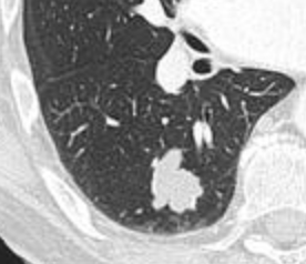
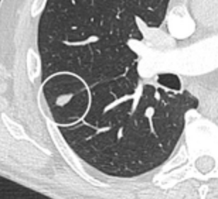
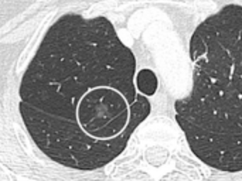
位置:上葉の位置は癌のリスク因子であり、 裂溝 や胸膜 に近い位置は良性リンパ節を示し、特に三角形の形状をしている場合はその可能性が高い。[ 8 ] [ 9 ] 辺縁 形態:棘状の辺縁は癌のリスク因子である。[ 8 ] [ 10 ] [ 9 ] [ 9 ] 境界が明瞭な円形の充実性肺結節で、滑らかな境界を持つ。
[ 9 ] 三角形の裂溝周囲リンパ節は良性リンパ節と診断されることがある。
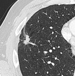
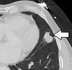
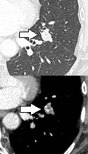
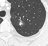
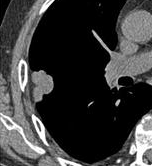
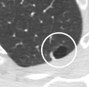
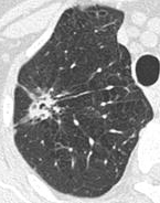
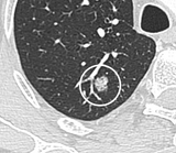
[ 9 ] 多発性 :最大3個の追加結節の存在は癌のリスクを高めることがわかっていますが、4個以上の追加結節の場合はリスクが低下します。これはおそらく癌ではなく、以前の肉芽腫性感染症を示しているためです。[ 8 ] 成長 率:固形がんは一般的に100~400日で体積が2倍になるのに対し、亜固形がん(一般的には腺癌)は一般的に3~5年で体積が2倍になる。[ 8 ] [ 8 ] 肺気腫 線維症 [ 8 ] [ 11 ] 造影効果:造影剤なしおよび 造影剤ありのCT 検査を併用した場合、造影効果が15ハウンズフィールド単位 (HU)未満の孤立性結節は悪性腫瘍を示唆するが、造影効果が高い場合は悪性腫瘍を示唆する(感度は 98%と推定される)。[ 12 ] 脂肪組織 の領域(-40~-120 HU)は過誤腫を 示唆する。しかし、過誤腫のうち脂肪を含むのは約50%にすぎない。[ 9 ] 中心空洞 がある場合、壁が薄い場合は良性疾患を示唆し、壁が厚い場合は悪性疾患を示唆する(特に4mm 以下と16mm 以上の場合)。[ 10 ] 石灰化とポップコーンのような外観から、過誤腫と診断される。[ 9 ] 石灰化 の場合、ポップコーンのような外観は過誤腫を示しており、これは良性である。[ 3 ] 亜実質性 結節の場合、一部が実質性である方が、純粋なすりガラス状陰影 である場合よりも癌のリスクが高い。Pleural retraction is far more common in cancers.[ 9] [ 9] Nodule with pleural retraction.
[ 9] In this case, pleural retraction is seen as a triangular fat component.
[ 9] Lung nodule abutting a pulmonary cyst.[ 9] A lung nodule abutting a pulmonary cyst is a rare finding, yet indicating cancer.[ 9] Bubble-like lucencies in the nodule indicate cancer:[ 9] Thin slice and maximal intensity projection of a lung nodule, the latter better visualizing vascular convergence.[ 9] Vascular convergence is where vessels converge to a nodule without adjoining or contacting the edge of the nodule, and is mainly seen in peripheral subsolid lung cancers.[ 9] angiogenesis .[ 9] Air bronchograms is defined as a pattern of air-filled bronchi on a background of airless lung, and may be seen in both benign and malignant nodules, but certain patterns thereof may help in risk stratification.[ 9]
CT densitometry, measuring absolute attenuation on the Hounsfield scale , has low sensitivity and specificity and is not routinely employed, apart from helping to distinguish solid from ground glass lesions, and to confirm visible fatty areas or calcifications.[ 12]
Diagnosis A diagnostic workup can include a variety of scans, blood tests, and biopsies.
Definition Nodular density is used to distinguish larger lung tumors, smaller infiltrates or masses with other accompanying characteristics. An often used formal radiological definition is the following: a single lesion in the lung completely surrounded by functional lung tissue with a diameter less than 3 cm and without associated pneumonia , atelectasis (lung collapse) or lymphadenopathies (swollen lymph nodes).[ 13] [ 10]
CT scan CTスキャンで偶発的に発見された結節については、Fleischner Societyの ガイドラインが以下の表に示されています。複数のリンパ節がある場合は、最も疑わしいリンパ節に基づいて管理されます。[ 8 ] [ 8 ]
推奨されているよりも頻繁にCTスキャンを行っても、結果が改善されることは示されておらず、放射線被曝が増加し、不必要な医療行為 によって患者が不安や不確実性を感じることが予想される。[ 14 ]
↑ Knipe, Henry. "コイン病変(肺)| 放射線学参考記事| Radiopaedia.org" . Radiopaedia . ↑ de Margerie-Mellon, Constance; Bankier, Alexander A. (2019年12月1日). "肺結節であるか否か..." . Radiology: Cardiothoracic Imaging . 1 (5) e190201. doi : 10.1148/ryct.2019190201 . PMC 7977753 . PMID 33778533 . 1 2 Ost D 、 Fein AM、Feinsilver SH ( 2003 年 6 月)。「臨床診療。孤立性肺結節」。 ニュー イングランド 医学ジャーナル 。348 (25): 2535–2542。doi : 10.1056/NEJMcp012290。PMID 12815140 。 1 2 3 Alzahouri K、Velten M、Arveux P、Woronoff-Lemsi MC、Jolly D、Guillemin F (2008 年 4 月)。 「フランスにおける SPN の 管理。孤立性肺結節の確定診断 の ための経路: フランス 18 地区における 多施設共同研究」 。BMC Cancer。8 : 93。doi : 10.1186 / 1471-2407-8-93。PMC 2373300。PMID 18402653 。 ↑ Thiessen NR、Bremner R ( 2010 年 10月) 。 「孤立性肺結節:一般外科医のアプローチ」。The Surgical Clinics of North America。90 ( 5): 1003–1018。doi : 10.1016 / j.suc.2010.07.002。PMID 20955880 。 1 2 Jude CM、Nayak NB、Patel MK、Deshmukh M、Batra P (2014)。「肺コクシジオイデス症:胸部X線およびCT所見の画像レビュー 」 Radiographics.34 ( 4 ) : 912–925.doi : 10.1148 / rg.344130134.PMID 25019431 . ↑ Zhan P, Xie H, Xu C, Hao K, Hou Z, Song Y (2013年12月). "孤立性肺結節の管理戦略" . Journal of Thoracic Disease . 5 (6): 824– 829. doi : 10.3978/j.issn.2072-1439.2013.12.13 . PMC 3886686 . PMID 24409361 . 1 2 3 4 5 6 7 8 9 10 11 MacMahon H、Naidich DP、Goo JM、Lee KS、Leung AN、Mayo JR、et al. (2017 年 7 月) 「CT 画像で検出された偶発性肺結節の管理に関するガイドライン: Fleischner Society 2017 より」 Radiology . 284 (1): 228– 243. doi : 10.1148/radiol.2017161659 . PMID 28240562 . 1 2 3 4 5 6 7 8 9 10 11 12 13 14 15 16 17 18 19 20 21 22 23 24 25 26 Snoeckx A、Reyntiens P、Desbuquoit D、Spinhoven MJ、Van Schil PE、van Meerbeeck JP、Parizel PM (2018 年 2 月)。 「孤立性肺結節 の評価: サイズ は重要 です が、形態の力を無視してはいけません」 。Insights into Imaging。9 ( 1 ): 73– 86。doi : 10.1007/ s13244-017-0581-2。PMC 5825309。PMID 29143191 。 1 2 3 Winer-Muram HT (2006 年 4 月) 「孤立性肺結節」 Radiology . 239 (1): 34– 49. doi : 10.1148/radiol.2391050343 . PMID 16567482 . ↑ Truong MT、Ko JP、Rossi SE、Rossi I、Viswanathan C、Bruzzi JF、et al . (2014 年 10 月) 。 「孤立 性 肺結節の評価の最新情報」。Radiographics。34 ( 6 ): 1658–1679。doi : 10.1148/rg.346130092。PMID 25310422 。 1 2 タナイ・パテル (2019-02-25)。 「肺転移画像診断」 。 メドスケープ 。 ↑ Tan BB、Flaherty KR 、 Kazerooni EA 、Iannettoni MD(2003 年1 月 )。 「 孤立性肺結節」 。Chest。123 (1 Suppl): 89S– 96S。doi : 10.1378 /chest.123.1_suppl.89S。PMID 12527568 。 {{cite journal}}: CS1メンテナンス: アーカイブサービスは非推奨になりました (リンク)↑ 米国胸部医師会 、 米国胸部学会 (2013年9月)。 「医師と患者が疑問に思うべき5つのこと」 。Choosing Wisely:ABIM財団のイニシアチブ 。米国胸部医師会および米国胸部学会。 2013年 1月6日 取得 。 Smith-Bindman R、Lipson J、Marcus R、Kim KP、Mahesh M、Gould R、et al. (2009 年 12 月) 「一般的なコンピュータ断層撮影検査に伴う放射線量と、それに関連する生涯がんリスク」 . Archives of Internal Medicine . 169 (22): 2078–2086 . doi : 10.1001/archinternmed.2009.427 . PMC 4635397 . PMID 20008690 . Wiener RS、Gould MK、Woloshin S、Schwartz LM、Clark JA(2013年3月)。「 スポットってどういう意味ですか?:肺結節について医師と話し合う患者の反応の定性的分析」 。Chest。143 ( 3 ) :672–677。doi :10.1378 / chest.12-1095。PMC 3590883。PMID 22814873 。 1 2 Gould MK、Maclean CC、Kuschner WG、Rydzak CE 、Owens DK(2001年2月)。「肺結節 および腫瘤性病変 の 診断 における陽電子放出断層撮影の精度:メタ分析」。JAMA。285 ( 7): 914–924。doi : 10.1001 / jama.285.7.914。PMID 11180735 。 ↑ Khan A (2007年3月)「孤立性肺結節に関するACRの適切性基準」 Journal of the American College of Radiology . 4 (3): 152– 155. doi : 10.1016/j.jacr.2006.12.003 . PMID 17412254 . ↑ Vansteenkiste JF、Stroobants SS(2006年1月)。「肺がんにおけるPETスキャン:現在の推奨事項と革新」。Journal of Thoracic Oncology。1 ( 1 ): 71–73。doi : 10.1097 / 01243894-200601000-00014。PMID 17409830 。 ↑ Cronin P、Dwamena BA、Kelly AM、Carlos RC(2008年3月)。「孤立性肺 結節:悪性腫瘍 の 診断のための断層画像診断法のメタアナリシス比較」。Radiology。246 ( 3 ) : 772–782。doi : 10.1148 / radiol.2463062148。PMID 18235105 。 ↑ Mukhopadhyay S (2012年1月) 「肺結節の診断における小生検の有用性:より少ないものでより多くのことを行う」 Modern Pathology . 25 (Suppl 1): S43– S57. doi : 10.1038/modpathol.2011.153 . PMID 22214970 . ↑ Klein JS、Salomon G、Stewart EA( 1996年3 月)。「同軸配置され た 20ゲージ自動切断針を用いた経胸壁針生検:122例の結果」。Radiology。198 ( 3 ) : 715–720。doi : 10.1148 /radiology.198.3.8628859。PMID 8628859 。 ↑ エラスムス JJ、マクアダムス HP、コノリー JE (2000)。 「孤立性肺結節: パート II. 不定形結節の評価」。 レントゲン写真 。 20 (1): 59–66 . doi : 10.1148/radiographics.20.1.g00ja0259 。 PMID 10682771 。