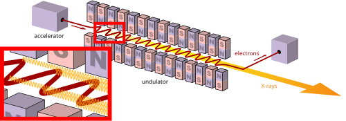
The radiation intensity grows, causing additional microbunching of the electrons, which continue to radiate in phase with each other.[9] This process continues until the electrons are completely microbunched and the radiation reaches a saturated power several orders of magnitude higher than that of the undulator radiation.
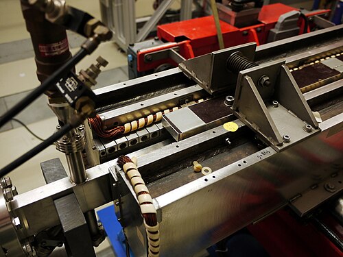
The wavelength of the radiation emitted can be readily tuned by adjusting the energy of the electron beam or the magnetic-field strength of the undulators.
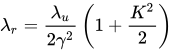
FELs are relativistic machines since the electrons driving the laser move at relativistic speeds—that is, at velocities very close to the speed of light. The wavelength of the emitted radiation, , is given by[10]
or when the wiggler strength parameter K, discussed below, is small
where is the undulator wavelength (the spatial period of the magnetic field), is the relativistic Lorentz factor and the proportionality constant depends on the undulator geometry and is of the order of 1.
This formula can be understood as a combination of two relativistic effects. Imagine you are sitting on an electron passing through the undulator. Due to Lorentz contraction the undulator is shortened by a factor and the electron experiences much shorter undulator wavelength . However, the radiation emitted at this wavelength is observed in the laboratory frame of reference and the relativistic Doppler effect brings the second factor to the above formula. In an X-ray FEL the typical undulator wavelength of 1cm is transformed to X-ray wavelengths on the order of 1nm by ≈ 2000, i.e. the electrons have to travel with the speed of 0.9999998c.
Wiggler strength parameter K
K, a dimensionless parameter, defines the wiggler strength as the relationship between the length of a period and the radius of bend,
where is the bending radius, is the applied magnetic field, is the electron mass, and is the elementary charge.
Expressed in practical units, the dimensionless undulator parameter is .
Quantum effects
In most cases, the theory of classical electromagnetism adequately accounts for the behavior of free electron lasers.[11] For sufficiently short wavelengths, quantum effects of electron recoil and shot noise may have to be considered.[12]
Construction
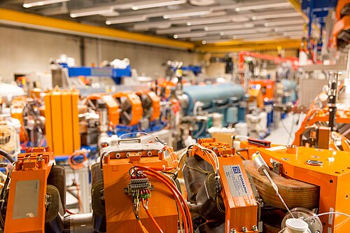
Free-electron lasers require the use of an electron accelerator with its associated shielding, as accelerated electrons can be a radiation hazard if not properly contained. These accelerators are typically powered by klystrons, which require a high-voltage supply. The electron beam must be maintained in a vacuum, which requires the use of numerous vacuum pumps along the beam path. While this equipment is bulky and expensive, free-electron lasers can achieve very high peak powers, and the tunability of FELs makes them highly desirable in many disciplines, including chemistry, structure determination of molecules in biology, medical diagnosis, and nondestructive testing.
At Helmholtz-Zentrum Dresden - Rossendorf two terahertz and mid-infrared FEL-based sources are in operation. FELBE is an FEL equipped with a cavity with continuous pulsing with a repetition rate of 13MHz, pulsing with 1kHz by applying a pulse picker, and macrobunch operation with bunch length > 100 μs and macrobunch repetition rates ≤ 25Hz. Pulse duration and pulse energy vary with wavelength and lie in the range from 1 - 25 ps and 100 nJ - few μJ, respectively.[15] The TELBE facility is based on a superradiant undulator offering THz pulses ranging from 0.1 THz to 2.5 THz at repetition rates up to 500kHz.[16]
X-ray FELs
The lack of mirror materials that can reflect extreme ultraviolet and x-rays means that X-ray free electron lasers (XFEL) need to work without a resonant cavity. Consequently, the beam is produced by a single pass of radiation through the undulator. This requires enough amplification over a single pass to produce a useful beam.
Exceptionally bright and fast X-rays can image proteins using x-ray crystallography. This technique allows first-time imaging of proteins that do not stack in a way that allows imaging by conventional techniques, 25% of the total number of proteins. Resolutions of 0.8nm have been achieved with pulse durations of 30 femtoseconds. To get a clear view, a resolution of 0.1–0.3nm is required. The short pulse durations allow images of X-ray diffraction patterns to be recorded before the molecules are destroyed.[29] The bright, fast X-rays were produced at the Linac Coherent Light Source at SLAC. As of 2014, LCLS was the world's most powerful X-ray FEL.[30]
Due to the increased repetition rates of the next-generation X-ray FEL sources, such as the European XFEL, the expected number of diffraction patterns is also expected to increase by a substantial amount.[31] The increase in the number of diffraction patterns will place a large strain on existing analysis methods. To combat this, several methods have been researched to sort the huge amount of data that typical X-ray FEL experiments will generate.[32][33] While the various methods have been shown to be effective, it is clear that to pave the way towards single-particle X-ray FEL imaging at full repetition rates, several challenges have to be overcome before the next resolution revolution can be achieved.[34][35]
One remaining challenge includes an efficient way to deliver single particles to the X-ray beam. Several different approaches are in development to address this issue, which includes electrospray ionization, gas-dynamic virtual nozzles and liquid sheet jets, which aim to provide stable and reproducible sample streams.[36][37]
It is of significance to consistently ensure that the particles intersect with the X-ray beam. As a result of introducing singular particles at a time, the hit rate on the beam is typically very low. This represents a major limitation with single-particle X-ray FEL imaging. Improvements to sample concentration and the precision of the delivery method is therefore crucial for increasing the efficiency of data collection.[38]
↑ Feldhaus, J.; Arthur, J.; Hastings, JB (2005). "X線自由電子レーザー" . Journal of Physics B . 38 (9): S799. Bibcode : 2005JPhB...38S.799F . doi : 10.1088/0953-4075/38/9/023 . S2CID 14043530 .
↑ Huang, Z.; Kim, K.-J. (2007). "X線自由電子レーザー理論のレビュー" . Physical Review Special Topics: Accelerators and Beams . 10 (3) 034801. Bibcode : 2007PhRvS..10c4801H . doi : 10.1103/PhysRevSTAB.10.034801 .
↑ Fain, B.; Milonni, PW (1987). "Classical stimulated emission". Journal of the Optical Society of America B . 4 (1): 78. Bibcode : 1987JOSAB...4...78F . doi : 10.1364/JOSAB.4.000078 .
↑ Benson, S.; Madey, JMJ (1984). "XUV自由電子レーザーにおける量子ゆらぎ". AIP Conference Proceedings . Vol. 118. pp. 173–182 . doi : 10.1063/1.34633 .
↑ MacKanos, MA; Joos, KM; Kozub, JA; Jansen, ED (2005). "パルス伸長自由電子レーザーを用いた角膜アブレーション". Manns, Fabrice; Soederberg, Per G; Ho, Arthur; Stuck, Bruce E; Belkin, Michael (編). Ophthalmic Technologies XV . Proceedings of the SPIE. Vol. 5688. p. 177. doi : 10.1117/12.596603 . S2CID 137024558 .
Elias, Luis R.; Fairbank, William M.; Madey, John MJ; Schwettman, H. Alan; Smith, Todd I. (1976). "空間的に周期的な横磁場における相対論的電子による誘導放出の観測" . Physical Review Letters . 36 (13): 717– 720. Bibcode : 1976PhRvL..36..717E . doi : 10.1103/physrevlett.36.717 .




![{\displaystyle K=0.934\cdot B_{0}\,{\text{[T]}}\cdot \lambda _{u}\,{\text{[cm]}}}](https://wikimedia.org/api/rest_v1/media/math/render/svg/c7c8af947de447492442fb88a51990081872b67f)