
Loading article…
Dental plaque is a biofilm of microorganisms (mostly bacteria, but also fungi) that grows on surfaces within the mouth. It is a sticky colorless deposit at first, but when it forms tartar, it is often brown or pale yellow. It is commonly found between the teeth, on the front of teeth, behind teeth, on chewing surfaces, along the gumline (supragingival), or below the gumline cervical margins (subgingival).[1] Dental plaque is also known as microbial plaque, oral biofilm, dental biofilm, dental plaque biofilm, and bacterial plaque biofilm. Bacterial plaque is one of the major causes for dental decay and gum disease.[1] It has been observed that differences in the composition of dental plaque microbiota exist between men and women, particularly in the presence of periodontitis.[2]
Progression and build-up of dental plaque can give rise to tooth decay, and periodontal problems. Hence it is important to disrupt the mass of bacteria and remove it.[3] Tooth decay is the localized destruction of the tissues of the tooth by acid produced from the bacterial degradation of fermentablesugar. Common periodontal problems include gingivitis and periodontitis.[4] Plaque control and removal can be achieved with correct daily or twice-daily tooth brushing and use of interdental aids such as dental floss and interdental brushes.[1]
Oral hygiene is important as dental biofilms may become acidic causing demineralization of the teeth (also known as dental caries) or harden into dental calculus (also known as tartar).[5] Calculus cannot be removed through tooth brushing or with interdental aids, but only through professional cleaning.[4]
歯垢は、放置すると歯の表面、修復物、および補綴装置(義歯やブリッジを含む)に付着するバイオフィルムです。歯垢の形成、構成、特性を理解することは、蓄積を抑制するのに役立ちます。 [ 6 ]獲得性ペリクルは、主に糖タンパク質で構成される唾液の層で、歯のクリーニング後または新しい歯が露出した直後に形成されます。[ 7 ]その後、細菌がペリクル層に付着し、微小コロニーを形成し、歯の上で成熟します。これが口腔疾患につながります。
口腔内には通常、さまざまな種類の細菌が存在します。これらの細菌は、白血球、好中球、マクロファージ、リンパ球とともに、正常な口腔内の一部であり、個人の健康に貢献しています。[ 1 ]歯垢の重量の約80~90%は水です。乾燥重量の70%は細菌であり、残りの30%は多糖類と糖タンパク質で構成されています。[ 8 ]
バイオフィルムを形成する微生物の大部分は、ミュータンス連鎖球菌とその他の嫌気性菌です。ただし、正確な構成は口腔内の場所によって異なります。そのような嫌気性菌の例としては、フソバクテリウム属と放線菌属が挙げられます。[ 1 ]ミュータンス連鎖球菌とその他の嫌気性菌は、歯の表面の最初の定着菌であり、初期のバイオフィルム群集の確立に重要な役割を果たします。[ 9 ]ミュータンス連鎖球菌は、グルカンスクラーゼという酵素を使用して、スクロースを粘着性のある細胞外のデキストランベースの多糖類に変換し、細菌が凝集してプラークを形成します。スクロースは、細菌がこの粘着性のある多糖類を形成するために使用できる唯一の糖です。[ 10 ]これらの微生物はすべて口腔内に自然に存在し、通常は無害です。しかし、定期的な歯磨きで歯垢を除去できないと、細菌は増殖し、厚い層となって蓄積し、宿主に様々な歯科疾患を引き起こす可能性があります。歯の表面に最も近い微生物は、通常、食事中のショ糖を発酵させてエネルギーを得ており、発酵の過程で酸を生成し始めます。
細菌の平衡状態は、プラーク形成の各段階で変化する。以下に、プラーク成熟の各段階で存在する可能性のある細菌の概要を示す。
歯垢は歯の表面に付着したバイオフィルムと考えられています。それは、特定の構造と機能に組織化された、精巧に形成された微生物群集です。[ 12 ]現代の技術を用いて約1000種類の細菌種が確認されていることから、歯垢は多様な種から構成されていると言えます。[ 13 ]
清潔な歯の表面はすぐに唾液ペリクルで覆われ、これが接着剤として機能します。これにより、最初の細菌(初期定着菌)が歯に付着し、定着して増殖することができます。初期定着菌がある程度増殖すると、バイオフィルムは後期定着菌と呼ばれる他の種類の細菌に対してより適合するようになります。[ 13 ]
出典: [ 13 ]
Source:[13]
Fusobacterium nucleatum is found between the early and late colonizers, linking them together.
Some salivary components are crucial for the plaque's ecosystem, such as salivary alpha-amylase which plays a role in binding and adhesion.[14] Proline-rich proteins (PRP) and statherins are also involved in the formation of plaque.[13]
Supragingival biofilm is dental plaque that forms above the gums, and is the first kind of plaque to form after brushing your teeth. It commonly forms in between the teeth, in the pits and grooves of the teeth, and along the gums. It is made up of mostly aerobic bacteria, meaning these bacteria need oxygen to survive. If plaque remains on the tooth for a long period of time, anaerobic bacteria begin to grow.[6]
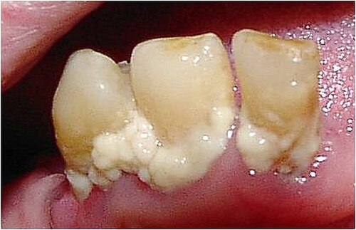
Subgingival biofilm is plaque that is located under the gums. It occurs after the formation of the supragingival biofilm by a downward growth of the bacteria from above the gums to below. This plaque is mostly made up of anaerobic bacteria, meaning that these bacteria will only survive if there is no oxygen. As this plaque attaches in a pocket under the gums, they are not exposed to oxygen in the mouth and will thrive if not removed.[11]
The extracellular matrix contains proteins, long-chain polysaccharides and lipids.
The most common reasons for ecosystem disruption are the ecological factors discussed in the environment section. The bacteria that exhibit the most fit plasticity for the change in environment dominate it. Often, this leads to opportunisticpathogens which may cause dental caries and periodontal disease. Pathogenic bacteria that have the potential to cause dental caries flourish in acidic environments; those that have the potential to cause periodontal disease flourish in a slightly alkaline environment.[15]
Antibodies to the oral pathogens Campylobacter rectus, Veillonella parvula, and Prevotella melaninogenica are associated with hypertension.[16]
Unlike other parts of the body, tooth surfaces are uniquely hard and non-shedding. Therefore, the warm and moist environment of the mouth and the presence of teeth, makes a good environment for growth and development of dental plaque.[13] The main ecological factors that contribute to plaque formation are pH, saliva, temperature and redox reactions.[17][18] The normal pH range of saliva is between 6 and 7 and plaque biofilm is known to flourish in a pH range between 6.7 and 8.3.[19][20] This indicates that the natural environment of the mouth provided by saliva is ideal for the growth of bacteria in the dental plaque. Saliva acts as a buffer, which helps to maintain the pH in the mouth between 6 and 7.[17] In addition to acting as a buffer, saliva and gingival crevicular fluid contain primary nutrients including amino acids, proteins and glycoproteins. This feeds the bacteria involved in plaque formation. The host diet plays only a minor role in providing nutrients for the resident microflora.[21] The normal temperature of the mouth ranges between 35 and 36 °C (95 and 97 °F), and a two-degree change has been shown to drastically shift the dominant species in the plaque.[18]Redox reactions are carried out by aerobic bacteria. This keeps the oxygen levels in the mouth at a semi-stable homeostatic condition, which allows the bacteria to survive.[18]

歯肉炎は、宿主と寄生体の相互作用によって引き起こされる炎症性病変で、歯肉組織に限局しています。これは、歯肉組織周辺にプラークが蓄積することによる一般的な結果です。バイオフィルム中に存在する細菌は、宿主の反応を引き起こし、組織の局所的な炎症につながります。[ 22 ]これは、歯肉の赤く腫れた外観や、ブラッシングやフロッシングによる出血など、炎症の主要な兆候によって特徴付けられます。[ 23 ]プラークによる歯肉炎は、プラークを除去することで回復できます。しかし、長期間放置すると、炎症が支持組織に影響を与え始め、歯周炎と呼ばれる進行をたどる可能性があります。[ 24 ] : 96–97歯肉炎反応は、多くの場合、歯周炎を回避する保護メカニズムです。
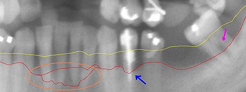
歯周炎は歯茎の感染症で、顎の歯の周囲の骨の破壊につながります。歯周炎は歯肉炎が確立された後に発生しますが、歯肉炎の人すべてが歯周炎になるわけではありません。[ 24 ] : 111 [ 25 ]歯垢の蓄積は歯周炎の進行に不可欠です。歯垢中の細菌が骨を攻撃して破壊する酵素を放出し、同時に骨内の破骨細胞がさらなる感染を防ぐ方法として骨を破壊します。これは、歯磨きや歯間の清掃などの厳格な口腔衛生、および歯科専門家による外科的デブリードメントによって治療できます。 [ 26 ]
歯垢による歯周炎の発症によって蓄積した細菌は、循環器系や呼吸器系を介して体内の遠隔部位に侵入し、様々な全身疾患や症状を引き起こす可能性があります。口腔内に存在する細菌は感染性があるため、細菌が体内に広がり、健康状態を悪化させる可能性があります。[ 27 ]細菌の侵入経路は、歯肉内の感染の蓄積によって生じる歯周ポケットの潰瘍化した上皮です。症状や疾患には以下のようなものがあります。
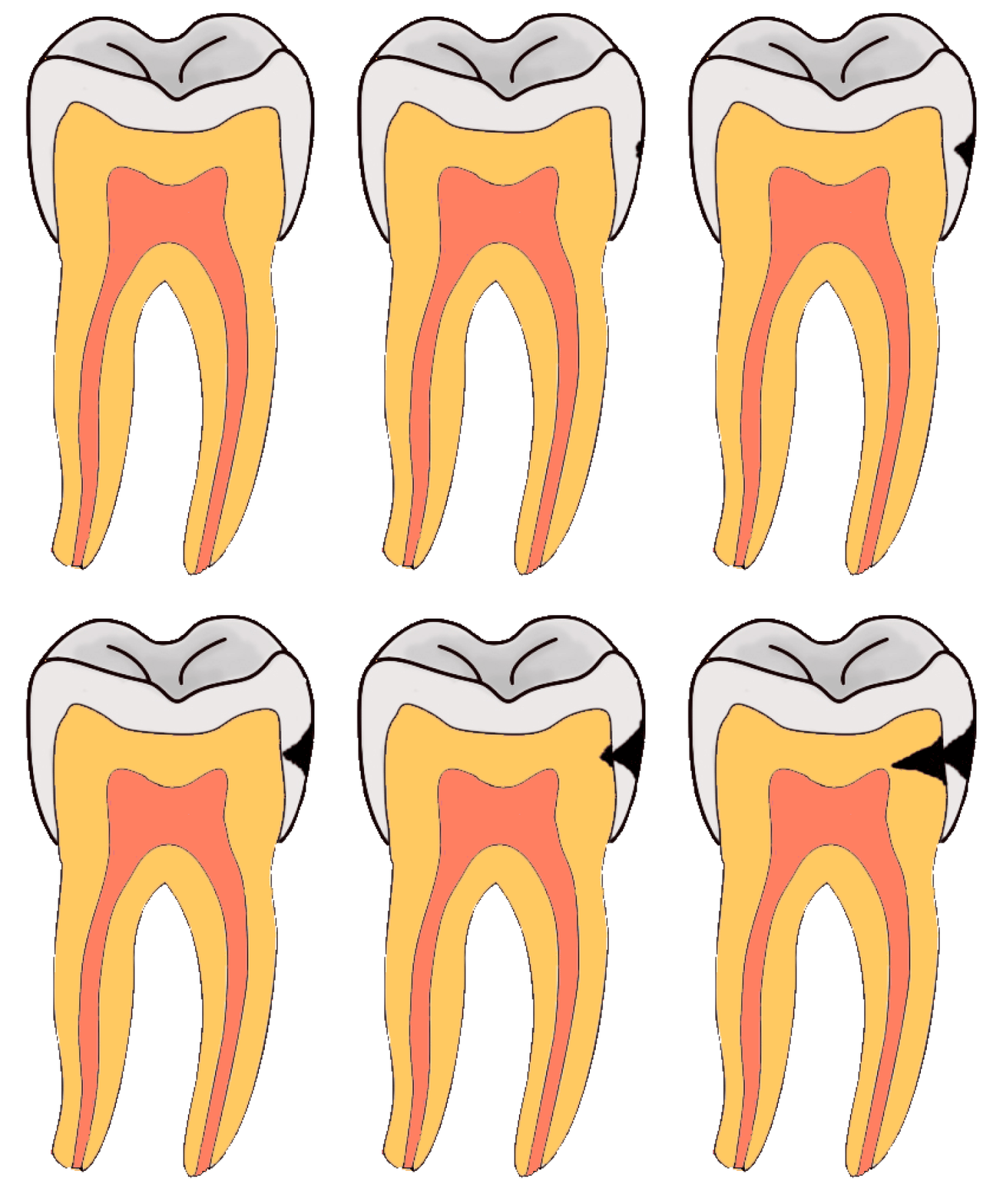
Dental caries is an infectious disease caused primarily by Streptococcus mutans, characterized by acid demineralization of the enamel, which can progress to further breakdown of the more organic, inner dental tissue (dentin).[1] The bacterial community would mainly consist of acidogenic and acid-tolerating species (e.g. Mutans streptococci and lactobacilli), while other species with relevant characteristics may also be involved.[32] Everybody is susceptible to caries but the probability of development depends on the patient's individual disease indicators, risk factors, and preventive factors. Factors that are considered high-risk for developing carious lesions on the teeth include:
Organic acids released from dental plaque lead to demineralization of the adjacent tooth surface, and consequently to dental caries. Saliva is also unable to penetrate the build-up of plaque and thus cannot act to neutralize the acid produced by the bacteria and remineralize the tooth surface.
There are two main methods of detecting dental plaque in the oral cavity: through the application of a disclosing gel or tablet, and/or visually through observation. Plaque detection is usually detected clinically by plaque disclosing agents. Disclosing agents contain dye which turns bright red to indicate plaque build-up.[1]
It is important for an individual to be aware of what to look for when doing a self-assessment for dental plaque. It is important to be aware that everyone has dental plaque, however, the severity of the build-up and the consequences of not removing the plaque can vary.[1]
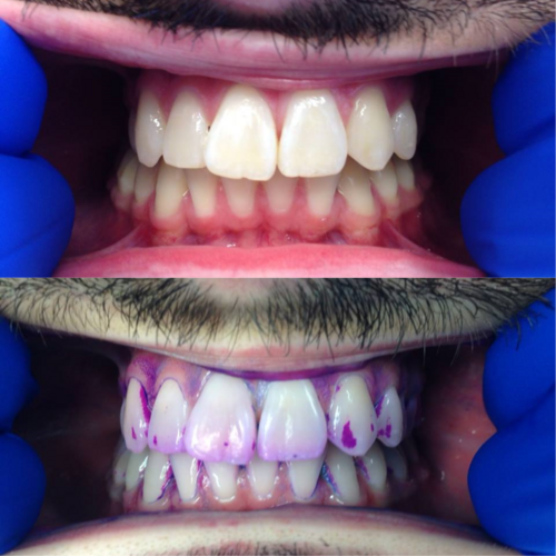
Plaque disclosing products, also known as disclosants, make plaque clinically visible. Clean surfaces of the teeth do not absorb the disclosant, only rough surfaces. Plaque disclosing gels can be completed either at home or in the dental clinic. Before using these at home or in the dental clinic check with your general practitioners for any allergies to iodine, food coloring, or any other ingredients that may be present in these products. These gels provide a visual aid in assessing plaque biofilm presence and can also show the maturity of the dental plaque.
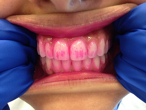
染色錠は染色ジェルと似ていますが、口に入れて約1分間噛む点が異なります。残った錠剤または唾液は吐き出します。染色ジェルは歯垢の存在を示しますが、歯垢の成熟度を示すことはあまりありません。染色錠は、最適な歯磨きを確実にするために、歯列矯正器具を装着している患者に、歯磨きの前後に使用するよう処方または渡されることがよくあります。また、歯垢の除去に苦労している幼児や患者にとって、歯垢除去の教育ツールとしても役立ちます。染色ジェルと染色錠は、あらゆる年齢層の人が効率的に歯垢を除去するのに役立ちます。
歯垢は、ブラッシング後わずか数分で歯に形成され始めます。歯垢は歯肉表面では見えにくいですが、ざらざらした表面として触ると分かります。多くの場合、厚く毛羽立ったような沈着物として感じられ、黄色、黄褐色、または茶色の染みとして現れることがあります。これらの沈着物は、歯や矯正器具などの歯科器具によく見られます。歯垢を評価する最も一般的な方法は、歯科医院での歯科検査で、歯科器具を使って歯垢を掻き取ることです。患者が歯垢を見つける最も一般的な場所は、歯と歯の間と歯頸部の縁です。
コンピュータ画像処理技術に関するあるレビューでは、歯垢の識別において高い精度が実証された。[ 33 ]いくつかの研究では、染色剤を使用しない場合、これが歯科医よりも優れた性能を発揮することが実証された。[ 33 ]
マウスウォッシュは、歯垢の蓄積を抑制する一般的な方法として用いられてきました。多くの研究で、アルコールを含むマウスウォッシュが最良の選択肢ではないことが示されています。アルコールを含むマウスウォッシュは、アルコールを含まないマウスウォッシュと比べて効果が大幅に高いわけではなく、アルコールを含むマウスウォッシュが口腔がんのリスクを高める可能性を示唆する証拠もいくつかあります。[ 34 ]マウスウォッシュにアルコールが含まれていないことから、多くのマウスウォッシュブランドが精油を使用した新しいマウスウォッシュを開発するようになりました。
2018年に、市販の精油入り洗口液の有効性に関する研究が行われた。プラセボとネガティブコントロールが用いられ、ネガティブコントロールは精油を含まない洗口液であった。健康なボランティア3グループに実験的に歯肉炎を誘発し、それぞれの洗口液を使用し、3週間観察した。結果は、精油入りの市販洗口液がプラークスコアにおいて有意に優れていることを示した。結果は、精油入り洗口液のプラークスコアが歯肉炎を予防するほど低くはなかったことを示した。研究者らは、精油入り洗口液の有効性は疑問であり、さらなる研究が必要であると結論付けた。[ 35 ]
2022年に行われた研究では、レモングラス精油の抗菌特性と効果が調査されました。 [ 36 ]実験室で歯垢の原因となる8種類の細菌が分離され、レモングラスオイルと接触させられました。その結果、レモングラスオイルは、別の口腔消毒薬であるクロルヘキシジンと同様に、これらの細菌の増殖を阻害することがわかりました。この研究では、レモングラス精油が実際に人間の歯垢形成を阻害するかどうかは調査されていません。
2022年に実施された研究では、209人の参加者を対象に、4種類の精油を混合したマウスウォッシュを使用した場合と、歯磨きとフロスのみを使用した場合の効果を比較しました。その結果、12週間後には、精油入りマウスウォッシュでうがいをしたグループは、歯磨きとフロスのみを行ったグループと比較して、歯垢が大幅に減少し、歯肉炎が改善したことが示されました。[ 37 ]
2021年に実施されたメタ分析では、さまざまなマウスウォッシュとその有効成分の歯垢に対する効果が検討されました。研究の収集には米国歯科医師会データベースが使用されました。概要のために合計22の論文が選択されました。選択された論文のうち4つ(すべてメタ分析)は、精油がかなりの抗歯垢活性を持つことを示しました。研究者らは、精油とクロルヘキシジンが口腔の健康維持に最も役立つ2つの成分であると結論付けました。[ 38 ]
20人の参加者を対象とした研究では、モクレン樹皮抽出物を含むマウスウォッシュは、ミュータンス連鎖球菌の発生率を減少させる点でプラセボよりも有意に優れていることがわかった。[ 39 ]
Dental plaque is extremely common in domestic animals such as dogs and cats. However, the bacteria associated with canine and feline plaque appear to be different from those in humans.[40][41] It consists of causing periodontal inflammation and triggers the animal's immune system. Two common distinctions that derive from periodontal are gingivitis which is inflammation, and periodontitis which involves the reactions of gingivitis causing severe gum disease. This severe gum disease can be something such as periodontitis and can ultimately lead to the loss of the tooth. Animals affected by periodontitis deal with irritation, and this disease can affect almost 80% of dogs above two years in age. Additionally, this disease is proven that with the focal point of gums, it can affect nearby organs.[42]
The periodontal disease is most attracted to the age and weight that the animals are currently in. The older and heavier they are the more likely they will catch the disease. A study of 9 female and 5 male dogs varying breeds and ages from 1-14 emphasizes the relation of Periodontal with age and no relation regarding breed or sex. A total of 50% of them were detected with the disease.[43] One study also mentioned that T. denticola was the most prevalent in dogs with periodontal disease and it was only located within affected dogs.[42]
The antibiotics that are commonly used for animals are antimicrobials ranging from clindamycin, amoxicillin-clavulate, and amoxicillin. These antimicrobials are commonly used in dental procedures for animals in the United States. A study was done were a total of 818,150 dogs and cats that resulted in the evident promotion of antimicrobials in retaining periodontitis. Other studies have been done that establish food debris as not being a concerning factor regarding the cause of dental plaque. In a 4-year study, a Beagle dog has been put to eat a strict diet without any oral hygiene which led to the accumulation of gingivitis in just a few weeks. However, some dogs were put on a similar diet with hygiene precautions of daily brushing their teeth and showed no signs of gingivitis.[44]
Animal owners have varying perspectives on what causes dental problems in their pets. Some believe that dry or moistened animal foods negatively impact dental health, while others believe that marrowbones can lead to fractures and discomfort or strengthen oral health.
Differences in breeds are also a familiar occurrence where owners disagree. Many perceive that distinctive breeds are more prone to health issues compared to others, while periodontitis is considered a smaller factor. Some owners express sentiments like "my dog has good teeth for their age," supporting the idea that as animals grow older, their dental features worsen, along with their weight.[45]
It is important to take care of pets by not only keeping them clean and providing them with healthy foods but also maintaining oral cleanliness to avoid discomfort and diseases. Hence, veterinarians often recommend oral healthcare products for affected pets.
