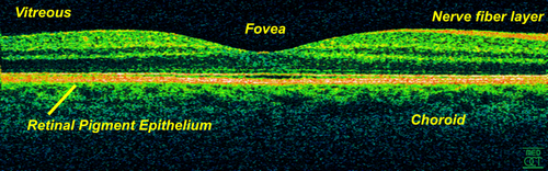
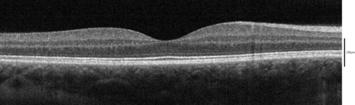
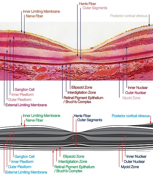
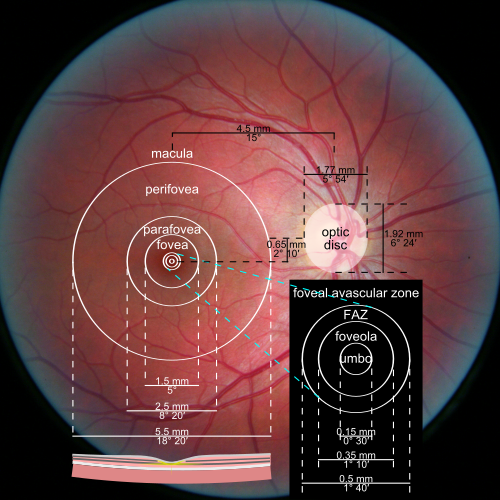
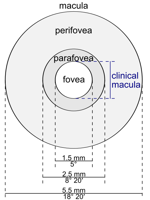
中心窩は網膜内表面のくぼみで、幅約 1.5 mm であり、その光受容体層はすべて錐体細胞で構成され、最高の視力に特化しています。中心窩内には、直径 0.5 mm の中心窩無血管帯(血管のない領域) と呼ばれる領域があります。これにより、光が分散したり失われたりすることなく感知されます。この解剖学的構造が中心窩の中央のくぼみの原因となっています。中心窩は、くぼみから移動したニューロンを含む中心窩縁に囲まれています。ここは網膜の中で最も厚い部分です。[ 6 ]
↑ Thibos, Larry; Lenner, Katharina; Thibos, Cameron (2023年12月18日). "Carl Bergmann (1814–1865) and the discovery of the anatomical site in the retina where vision is starteded" . Journal of the History of the Neurosciences . 33 (2): 180– 203. doi : 10.1080/0964704X.2023.2286991 . PMID 38109332 . S2CID 266361309 .
↑ Provis, Jan M; Dubis, Adam M; Maddess, Ted; Carroll, Joseph (2013). "Adaptation of the central retina for high acuity vision: Cones, the fovea and the avascular zone" . Progress in Retinal and Eye Research . 35 : 63–81 . doi : 10.1016/j.preteyeres.2013.01.005 . PMC 3658155. PMID 23500068 .
↑ Tschulakow, Alexander V; Oltrup, Theo; Bende, Thomas; Schmelzle, Sebastian; Schraermeyer, Ulrich (2018). " The anatomy of the foveola reinvestigated" . PeerJ . 6 e4482. doi : 10.7717/peerj.4482 . PMC 5853608. PMID 29576957 .この資料は、クリエイティブ・コモンズ表示4.0国際ライセンスの下で利用可能なこの情報源からコピーされました。
↑ Foulsham, Tom; Walker, Esther; Kingstone, Alan (2011). "The where, what and when of gaze allocation in the lab and the natural environment" . Vision Research . 51 (17): 1920–1931 . doi : 10.1016/j.visres.2011.07.002 . ISSN 0042-6989 . PMID 21784095. S2CID 17511680 .
↑ Sailer, U. (2005-09-28). "新規視覚運動課題の学習中の目と手の協調" . Journal of Neuroscience . 25 (39): 8833– 8842. doi : 10.1523/jneurosci.2658-05.2005 . ISSN 0270-6474 . PMC 6725583 . PMID 16192373 .
↑ Krinsky, Norman I; Landrum, John T; Bone, Richard A (2003). "眼におけるルテインとゼアキサンチンの保護的役割の生物学的メカニズム". Annual Review of Nutrition . 23 : 171–201 . doi : 10.1146/annurev.nutr.23.011702.073307 . PMID 12626691 .
↑ Landrum, John T; Bone, Richard A (2001). "ルテイン、ゼアキサンチン、黄斑色素". Archives of Biochemistry and Biophysics . 385 (1): 28– 40. doi : 10.1006/abbi.2000.2171 . PMID 11361022 .
↑ Beatty, S; Boulton, M; Henson, D; Koh, HH; Murray, IJ (1999). "黄斑色素と加齢黄斑変性" . British Journal of Ophthalmology . 83 (7): 867– 877. doi : 10.1136/bjo.83.7.867 . PMC 1723114 . PMID 10381676 .
1 2 Curcio, Christine A; Allen, Kimberly A; Sloan, Kenneth R; Lerea, Connie L; Hurley, James B; Klock, Ingrid B; Milam, Ann H (1991). "抗ブルーオプシンで染色したヒト錐体光受容体の分布と形態". The Journal of Comparative Neurology . 312 (4): 610– 624. doi : 10.1002/cne.903120411 . PMID 1722224 . S2CID 1947541 .
↑ Serpenguzel, Ali; Serpengüzel, Ali; Poon, Andrew W. (2011). Optical Processes in Microparticles and Nanostructures: A Festschrift Dedicated to Richard Kounai Chang on His Retirement from Yale University . World Scientific. ISBN978-981-4295-77-2。
↑ Curcio, Christine A; Sloan, Kenneth R; Kalina, Robert E; Hendrickson, Anita E (1990). "ヒト光受容体トポグラフィー". The Journal of Comparative Neurology . 292 (4): 497– 523. doi : 10.1002/cne.902920402 . PMID 2324310 . S2CID 24649779 .
↑ Vannasdale, D. A; Elsner, A. E; Weber, A; Miura, M; Haggerty, B. P (2009). "走査型レーザー偏光測定法を用いた中心窩位置の決定" . Journal of Vision . 9 (3): 21.1–17. doi : 10.1167/9.3.21 . PMC 2970516 . PMID 19757960 .
1 2 Magnussen, Svein ; Spillmann, Lothar; Stürzel, Frank; Werner, John S (2001). "中心窩青色暗点の塗りつぶし" . Vision Research . 41 (23): 2961– 2967. doi : 10.1016/S0042-6989(01)00178-X . PMC 2715890 . PMID 11704235 .
↑ Flom, M. C; Weymouth, F. W (1961). "斜視と弱視におけるマクスウェル斑の中心性". Archives of Ophthalmology . 66 (2): 260– 268. doi : 10.1001/archopht.1961.00960010262018 . PMID 13700314 .



 この資料は、クリエイティブ・コモンズ表示4.0国際ライセンスの下で利用可能なこの情報源からコピーされました。