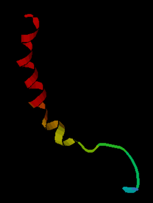
Neuropeptides are synthesized from inactive precursor proteins called prepropeptides.[7] Prepropeptides contain sequences for a family of distinct peptides and often contain duplicated copies of the same peptides, depending on the organism.[8] In addition to the precursor peptide sequences, prepropeptides also contain a signal peptide, spacer peptides, and cleavage sites.[9] The signal peptide sequence guides the protein to the secretory pathway, starting at the endoplasmic reticulum. The signal peptide sequence is removed in the endoplasmic reticulum, yielding a propeptide. The propeptide travels to the Golgi apparatus where it is proteolytically cleaved and processed into multiple peptides. Peptides are packaged into dense core vesicles, where further cleaving and processing, such as C-terminal amidation, can occur. Dense core vesicles are transported throughout the neuron and can release peptides at the synaptic cleft, cell body, and along the axon.[7][10][11][12]
A single animal may use hundreds of different neuropeptides. In C. elegans, for example, 120 genes specify more than 250 neuropeptides.[13]
Mechanism
Neuropeptides are released by dense core vesicles after depolarization of the cell. Compared to classical neurotransmitter signaling, neuropeptide signaling is more sensitive. Neuropeptide receptor affinity is in the nanomolar to micromolar range while neurotransmitter affinity is in the micromolar to millimolar range. Additionally, dense core vesicles contain a small amount of neuropeptide (3 - 10mM) compared to synaptic vesicles containing neurotransmitters (e.g. 100mM for acetylcholine).[14] Evidence shows that neuropeptides are released after high-frequency firing or bursts, distinguishing dense core vesicle from synaptic vesicle release.[10] Neuropeptides utilize volume transmission and are not reuptaken quickly, allowing diffusion across broad areas (nm to mm) to reach targets. Almost all neuropeptides bind to G protein-coupled receptors (GPCRs), inducing second messenger cascades to modulate neural activity on long time-scales.[7][10][11]
↑ Jékely G (2021年3月) 「神経系の起源に関する化学的脳仮説」 . Philosophical Transactions of the Royal Society of London. Series B, Biological Sciences . 376 (1821) 20190761. doi : 10.1098/rstb.2019.0761 . PMC 7935135 . PMID 33550946 .
↑Sachkova MY, Nordmann EL, Soto-Àngel JJ, Meeda Y, Górski B, Naumann B, etal. (December 2021). "Neuropeptide repertoire and 3D anatomy of the ctenophore nervous system". Current Biology. 31 (23): 5274–5285.e6. Bibcode:2021CBio...31E5274S. doi:10.1016/j.cub.2021.09.005. PMID34587474. S2CID238210404.
↑Takahashi T, Takeda N (January 2015). "Insight into the molecular and functional diversity of cnidarian neuropeptides". International Journal of Molecular Sciences. 16 (2): 2610–2625. doi:10.3390/ijms16022610. PMC4346854. PMID25625515.
↑Mirabeau O, Joly JS (May 2013). "Molecular evolution of peptidergic signaling systems in bilaterians". Proceedings of the National Academy of Sciences of the United States of America. 110 (22): E2028–E2037. Bibcode:2013PNAS..110E2028M. doi:10.1073/pnas.1219956110. PMC3670399. PMID23671109.
↑Yañez-Guerra LA, Thiel D, Jékely G (April 2022). "Premetazoan Origin of Neuropeptide Signaling". Molecular Biology and Evolution. 39 (4) msac051. doi:10.1093/molbev/msac051. PMC9004410. PMID35277960.