
Loading article…
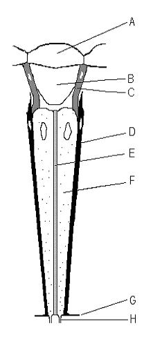

昆虫、甲殻類、ヤスデなどの節足動物の複眼[ 1 ]は、個眼(単数形:オマチジウム)と呼ばれる単位で構成されています。オマチジウムには、支持細胞と色素細胞に囲まれた光受容細胞の集まりが含まれています。オマチジウムの外側は透明な角膜で覆われています。各オマチジウムは1つの軸索束(通常、桿状体数に応じて6~9本の軸索から構成される)[ 2 ] : 162によって神経支配され、脳に1つの画像要素を提供します。脳はこれらの独立した画像要素から画像を形成します。眼の個眼の数は節足動物の種類によって異なり、南極等脚類のGlyptonotus antarcticus [ 3 ]のようにわずか 5 個、原始的なZygentomaのように一握りのものから、大型のAnisopteraトンボや一部のSphingidaeガのように約 30,000 個まで様々である。[ 4 ]
個眼は通常、断面が六角形で、長さは幅の約10倍である。直径は表面で最も大きく、内側に向かって細くなっている。外表面には角膜があり、その下には光をさらに集束させる偽円錐がある。角膜と偽円錐は個眼の長さの外側10%を占める。
個眼、つまり眼の単位の具体的な器官は、生物によって異なります。チョウの複眼は複数の個眼からなり、それぞれの個眼は9つの光受容細胞(R1~R9と番号付けされている)と一次および二次色素細胞から構成されています。[ 5 ]タテハチョウ科のチョウは最も単純な眼の個眼構造を持ち、8つの光受容細胞(R1~R8)と、別の階層に配置された小さなR9細胞から構成されています。[ 5 ]これらの「R細胞」は個眼を密に詰め込んでいます。個眼の中心軸にあるR細胞の部分は集合的に光導波路、つまり桿状体と呼ばれる透明な管を形成します。
ショウジョウバエの複眼は16,000個以上の細胞から構成されているが[ 6 ] 、幼虫の眼原基で形成される700~750個の個眼が単純に繰り返し配列した構造である[ 7 ] 。各個眼は14個の隣接する細胞から構成されており、中心部には8個の光受容ニューロン、4個の非神経性錐体細胞、2個の一次色素細胞がある[ 6 ] 。色素細胞の六角形の格子が個眼の中心部を隣接する個眼から隔離し、視野のカバー範囲を最適化することで、ショウジョウバエの視力の鋭さに影響を与える[ 7 ]。
真のハエでは、桿状体は 7 つの独立した桿状体節に分かれており (実際には 8 つあるが、色覚を司る 2 つの中央の桿状体節が上下に重なっている)、各個眼に 7 ピクセルの小さな反転画像が形成されます。 単一の個眼内の隣接する桿状体節間の角度 (受容角) は、隣接する個眼間の角度 (個眼間角度) と似ているため、隣接する個眼間に重なり合う領域を持つ連続した視野が眼に与えられます。[ 2 ] : 161–2この配置の利点は、同じ視軸が眼のより広い領域からサンプリングされるため、眼のサイズを大きくしたり、視力を低下させたりすることなく、全体的な感度が 7 倍に増加することです。これを実現するには、眼の配線を再構築する必要があり、軸索束を180度ねじり(反転させ)、各桿状節を同じ視軸を共有する6つの隣接する個眼の桿状節と結合させる必要があった。したがって、昆虫の脳の最初の光学処理センターであるラミナのレベルでは、信号は通常の対向型複眼の場合とまったく同じ方法で入力されるが、画像は強調される。この視覚配置は神経重ね合わせとして知られている。[ 2 ]: 163-4
Since an image from the compound eye is created from the independent picture elements produced by ommatidia, it is important for the ommatidia to react only to that part of the scene directly in front of them. To prevent light entering at an angle from being detected by the ommatidium it entered, or by any of the neighboring ommatidia, six pigment cells are present. The pigment cells line the outside of each ommatidium. Pigment cells are located at an ommatidium's vertices, thus each pigment cell lines the outside of three mutually-adjacent ommatidia. Light entering at an angle passes through the thin cross-section of the photoreceptor cell, with only a tiny chance of exciting it, and is absorbed by the pigment cell, before it can enter a neighboring ommatidium. In many species, in low-light situations, the pigment is withdrawn, so that light entering the eye might be detected by any of several ommatidia. This enhances light detection but lowers resolution.
The size of the ommatidia varies according to species, but ranges from 5 to 50 micrometres. The rhabdoms within them may cross-section at least as small as 1.x micrometres, the category of "small" being assigned in some cross-species studies to those under 2 micrometers.[8] A microlens array can be seen as an elementary, biomimetic analogy of ommatidia.
Retinal cell fate determination relies on positional cell–cell signaling that activates signal transduction pathways, rather than cell lineage. Cell–cell signals released from R8 photoreceptors (already differentiated retinal cells) of each ommatidium are received by neighboring progenitor retinal cells, stimulating their incorporation into developing ommatidia. The undifferentiated retinal cells select their appropriate cell fates based on their position with their differentiated neighbors. The local signal, Growth Factor Spitz, activates the epidermal growth factor receptor (EGFR) signal transduction pathway, and initiates a cascade of events that will result in transcription of genes involved in cell fate determination.[9] This process leads to the induction of cell fates, starting from the R8 photoreceptor neurons and progresses to the sequential recruitment of neighboring undifferentiated cells. The first seven neighboring cells receive R8 signaling to differentiate as photoreceptor neurons, followed by the recruitment of the four non-neuronal cone cells.