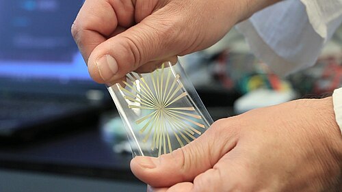
伸縮性マイクロ電極アレイ(伸縮性MEAまたはsMEA) (伸縮性マルチ電極アレイとも呼ばれる)は、特殊なタイプのマイクロ電極アレイ(MEA)であり、重要な利点があります。それは、標準的なMEAが機械的負荷によって破損するのに対し、伸縮性MEAは電気的機能を維持したまま変形、伸張、曲げ、ねじりができることです。伸縮性MEAと混同されがちなフレキシブルMEA(flexMEA)は、ある程度曲げたりねじったりすることはできますが、伸張はしないため、機械的特性の点では伸縮性MEAと標準MEAの中間に位置します。従来のMEAと同様に、伸縮性MEAは、細胞(ニューロン、筋肉など)からの電気信号の記録または刺激を可能にする数千個のマイクロ電極で構成されており、生体内でのin vivo 、または細胞培養を用いたin vitroで使用されます。
Geometric patterning, fractal patterns: Metal traces are deposited in specific patterns, such as meandering or serpentine shapes, within a stretchable elastomeric substrate to accommodate strain. The resulting structure is akin to a 2-dimensional spring. The University of Ghent and IMEC in Belgium have pioneered the approach to using Meander shaped metallic structures.[8]
The group of John Rogers increased the maximum strain in devices created by this approach using fractal-based structures. These fractal patterns are characterized by self-similarity, i.e., a small sections of the structure yields pieces with geometries that resemble the whole structure.
These fractal patterns include (i) Koch, Peano, Hilbert lines, (ii) Moore, Vicsek loops, and (iii) Greek crosses.[9]
Origami-inspired structures, and kirigami cuts: Intrinsically rigid or inelastic flexible materials can be turned into stretchable materials by applying origami technology[10] and kirigami cuts.[11]
History
The first time the term stretchable multielectrode array (sMEA)
Manually stretching sMEA
Understanding how cells convert mechanical stimuli appeared in the literature was in a conference proceeding in 2002 from the Lawrence Livermore National Laboratory.[12] This paper described the fabrication of an sMEA for a retinal prosthesis, but no biological material was used, i.e., functionality to record or stimulate neural activity was not attempted. The first description of sMEAs being used to record neural activity in biological samples was in 2006 when the research group of Barclay Morrison at Columbia University and Sigurd Wagner at Princeton University reported recording of spontaneous activity in organotypic hippocampal tissue slices.[13] Neither the electrodes nor the tissue appears to have been stretched in these experiments. In 2008, a paper from the Georgia Institute of Technology and Emory University described the use of sMEAs in stimulating a explant of a rat spinal cord.[14] The sMEA was wrapped around the spinal cord, but not stretched, and the cells were electrically stimulated but not used in recording electrophysiological activity. In 2009, another paper of the Morrison/Wagner groups described for the first time the use of an sMEA with a biological sample being stretched as well as electrical stimulation and recording of electrophysiological activity being carried out before and after stretching.[15]
In subsequent years, the number of research papers that describes different approaches to fabricating sMEAs and their use for in vitro and in vivo research has increased immensely.
Types and capabilities
Stretchable microelectrode arrays (sMEAs) can be categorized whether they are used with cells or tissue slices in a dish (in vitro) or whether they are implanted in an animal or human (in vivo).
In vitro stretchable MEAs
sMEAs are used in vitro to record and stimulate electrophysiological activity in dissociated cells, tissue slices or organoids. In vitro use of sMEAs may include stretching of the cells. The cells are either harvested from an animal or were derived from human induced pluripotent stem cells (hiPSCs).
The form factor of sMEAs is often similar to rigid MEAs because the same data acquisition systems can be used for both types of MEAs. The main differences between sMEAs and rigid MEAs are summarized below:
The main disadvantage of sMEAs compared to rigid MEAs are related to the different technologies that are used to manufacture these devices. sMEAs have usually up to 60 electrodes with diameters of between 50μm and 100μm where rigid CMOS based MEAs can have thousands of electrodes with diameters of 10μm. This means that sMEAs are not suitable for studying sub-cellular structures.
In vivo stretchable MEAs
Stretchable MEAs have many benefits for implantable in vivo applications for recording and stimulation of electrophysiological activity from electrogenic biological tissues (most commonly neurons and muscles). Some applications involve only recording of electrophysiological activity, e.g., on the surface of the brain,[18] the spinal cord,[19] some involve only stimulation of electrophysiological activity, and some both.[20]
Advantages
The main benefits of using sMEAs for in vivo applications are twofold. First, they can conform to the dynamic and often curved surfaces of biological tissues. Second, sMEAs cause significant smaller foreign body reaction than rigid MEAs because of the reduced mismatch in mechanical properties (stiffness) between the implant the tissue.[21]
Disadvantage
The main disadvantage of sMEAs for implanted applications is the mechanical robustness compared to rigid MEAs, which can cause the implant to break or tear.
Applications
Neural interfaces
In neural interfaces, sMEAs are utilized to record and stimulate neural activity. Their stretchability allows them to conform to the brain's surface or penetrate neural tissue without causing significant damage.[21] This improves the quality of neural recordings and the effectiveness of neural stimulation, which is crucial for applications such as brain-machine interfaces.[21]
Electrocorticography
Electrocorticography (EcoG) with stretchable MEAs offers a less invasive method for recording electrical activity from the brain's surface. These arrays can conform to the cortical surface, providing high-resolution, stable recordings even during brain movements. This capability is essential for applications such as epilepsy monitoring and brain-computer interfaces.
↑ Kyrylyuk, Andriy V.; van der Schoot, Paul (2008年6月17日). "ポリマーおよびコロイド媒体におけるカーボンナノチューブの連続体パーコレーション" . Proceedings of the National Academy of Sciences . 105 (24): 8221– 8226. Bibcode : 2008PNAS..105.8221K . doi : 10.1073/pnas.0711449105 . PMC 2448818 . PMID 18550818 .
↑ Pionteck, Jürgen; Wypych, George 編 (2016). 「非移動性帯電防止剤の構造と分布」『帯電防止剤ハンドブック』pp. 117–127 . doi : 10.1016/B978-1-895198-95-9.50011-X . ISBN978-1-895198-95-9。
↑ Huck, Wilhelm TS; Bowden, Ned; Onck, Patrick; Pardoen, Thomas; Hutchinson, John W.; Whitesides, George M. (2000 年 4 月). "平面表面上の自然発生的な座屈の秩序化". Langmuir . 16 (7): 3497– 3501. doi : 10.1021/la991302l .
↑ Kim, Dae-Hyeong; Rogers, John A. (2008年12月17日). "伸縮性エレクトロニクス:材料戦略とデバイス". Advanced Materials . 20 (24): 4887–4892 . Bibcode : 2008AdM....20.4887K . doi : 10.1002/adma.200801788 .
↑ Maghribi, M.; Hamilton, J.; Polla, D.; Rose, K.; Wilson, T.; Krulevitch, P. (2002). "伸縮性マイクロ電極アレイ[網膜プロテーゼ用] ".第2回IEEE-EMBS国際マイクロテクノロジー医学・生物学特別トピック会議議事録(カタログ番号02EX578) . pp. 80–83 . doi : 10.1109/MMB.2002.1002269 . ISBN0-7803-7480-0。
↑ Yu, Zhe; Tsay, Candice; Lacour, Stephanie P.; Wagner, Sigurd; Morrison, Barclay (2006). "伸縮性マイクロ電極アレイ:外傷性脳損傷の根底にある機能障害のメカニズムの発見と神経細胞と神経補綴のインターフェースのためのツール". 2006 International Conference of the IEEE Engineering in Medicine and Biology Society . Vol. Suppl. pp. 6732–6735 . doi : 10.1109/IEMBS.2006.260933 . ISBN1-4244-0032-5PMID 17959498 .
↑ Meacham, Kathleen W.; Giuly, Richard J.; Guo, Liang; Hochman, Shawn; DeWeerth, Stephen P. (2008 年 4 月). "脊髄表面刺激のためのリソグラフィーパターン化された弾性多電極アレイ" . Biomedical Microdevices . 10 (2): 259– 269. doi : 10.1007/s10544-007-9132-9 . PMC 2573864 . PMID 17914674 .