
Loading article…
In biology, depolarization or hypopolarization[1][2] is a change within a cell, during which the cell undergoes a shift in electric charge distribution, resulting in less negative charge inside the cell compared to the outside. Depolarization is essential to the function of many cells, communication between cells, and the overall physiology of an organism. It is especially important to electrical signaling in neurons and muscle cells. It also affects many non-excitable cells by changing calcium regulation or gene expression.[3]
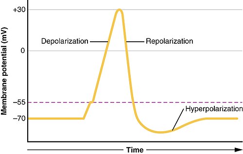
Most cells in higher organisms maintain an internal environment that is negatively charged relative to the cell's exterior. This difference in charge is called the cell's membrane potential. In the process of depolarization, the negative internal charge of the cell temporarily becomes more positive (less negative). This shift from a negative to a more positive membrane potential occurs during several processes, including an action potential. During an action potential, the depolarization is so large that the potential difference across the cell membrane briefly reverses polarity, with the inside of the cell becoming positively charged.
The change in charge typically occurs due to an influx of sodiumions into a cell, although it can be mediated by an influx of any kind of cation or efflux of any kind of anion. The opposite of a depolarization is called a hyperpolarization.
Usage of the term "depolarization" in biology differs from its use in physics, where it refers to situations in which any form of electrical polarity (i.e. the presence of any electrical charge, whether positive or negative) changes to a value of zero.
Depolarization is sometimes referred to as "hypopolarization"[1][2] (as opposed to hyperpolarization).
The process of depolarization is entirely dependent upon the intrinsic electrical nature of most cells. When a cell is at rest, the cell maintains what is known as a resting potential.[4] The resting potential generated by nearly all cells results in the interior of the cell having a negative charge compared to the exterior of the cell.[4] To maintain this electrical imbalance, ions are transported across the cell's plasma membrane.[5] The transport of the ions across the plasma membrane is accomplished through several different types of transmembrane proteins embedded in the cell's plasma membrane that function as pathways for ions both into and out of the cell, such as ion channels, sodium potassium pumps, and voltage-gated ion channels.[6]
The resting potential must be established within a cell before the cell can be depolarized. There are many mechanisms by which a cell can establish a resting potential, however there is a typical pattern of generating this resting potential that many cells follow. The generation of a negative resting potential within the cell involves the utilization of ion channels, ion pumps, and voltage-gated ion channels by the cell.[7] However, the process of generating the resting potential within the cell also creates an environment outside the cell that favors depolarization. The sodium potassium pump is largely responsible for the optimization of conditions on both the interior and the exterior of the cell for depolarization. This pump is an ATPase that pumps three sodium ions out of the cell and two potassium ions into the cell with every ATP consumed.[8]
細胞内に2つの正電荷を持つカリウムイオン(K + )が送り込まれるごとに、3つの正電荷を持つナトリウムイオン(Na +)が細胞外に送り出されることで、各ATP分子で+1の電荷が失われるため細胞内部の正電荷が小さくなり、細胞の静止電位が確立されるだけでなく、細胞外のナトリウム濃度と細胞内のカリウム濃度が増加するため、好ましくない濃度勾配が作られます。 [ 9 ]細胞内に過剰なカリウムと細胞外のナトリウムが存在する間、生成された静止電位は、形質膜の電圧依存性イオンチャネルの閉鎖を維持します。これは、膜を横切って送り込まれたイオンの拡散を防ぐだけでなく、カリウムリークチャネルの活動も関与し、カリウムイオンの制御された受動的な流出を可能にし、負の静止電位の確立に寄与します。[ 10 ]さらに、高濃度の正電荷を持つカリウムイオンにもかかわらず、ほとんどの細胞には(負電荷を持つ)内部成分が含まれており、それが蓄積して負の内部電荷を確立します。
細胞が静止電位を確立した後、その細胞は脱分極を起こす能力を持つ。脱分極とは、膜電位がより負の電位から負の電位へと変化し、活動電位の発生を促進する過程である。[ 10 ]細胞内部でこのような急速な変化が起こるためには、細胞の形質膜に沿っていくつかの事象が発生する必要がある。
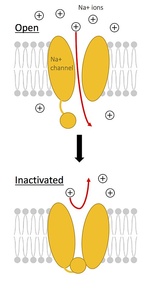
ナトリウム-カリウムポンプが働き続ける一方で、細胞が静止電位にある間は閉じていた電位依存性ナトリウムチャネルとカルシウムチャネル[ 11 ]は、最初の電圧変化に応じて開きます。 [ 10 ]ニューロンの電荷の変化が電位依存性ナトリウムチャネルの開口につながると、電気化学的勾配に従ってナトリウムイオンが流入します。ナトリウムイオンは細胞内に入り、細胞内部に正の電荷を与え、膜電位を負から正に変化させます。最初のナトリウムイオン流入は、追加のナトリウムチャネルの開口(正のフィードバックループ)を引き起こし、細胞内へのナトリウムイオンのさらなる移動につながり、正の平衡電位に達するまで脱分極プロセスを維持します。[ 12 ]
ナトリウムチャネルは、膜が脱分極状態であっても、迅速な再閉鎖を促す固有の不活性化機構を備えている。この平衡状態では、ナトリウムチャネルは不活性化状態に入り、膜電位が再び負に帯電するまでナトリウムイオンの流入を一時的に停止する。細胞内部が十分に正に帯電すると、脱分極が終了し、チャネルは再び閉じる。[ 10 ]
細胞が脱分極した後、細胞内部の電荷に最後の変化が生じます。脱分極後、脱分極中に開いていた電位依存性ナトリウムイオンチャネルは再び閉じます。細胞内の正電荷の増加により、カリウムチャネルが開きます。カリウムイオン(K +)は電気化学的勾配(濃度勾配と新たに確立された電気勾配に有利な方向)に沿って移動し始めます。カリウムが細胞外に移動するにつれて、細胞内の電位は低下し、再び静止電位に近づきます。ナトリウムカリウムポンプはこのプロセス全体を通して継続的に機能します。[ 13 ]
出典:[ 3 ]
再分極の過程で細胞電位がオーバーシュートする。カリウムイオンは軸索から移動し続け、静止電位を超え、新しい細胞電位は静止電位よりも負になる。最終的に、すべての電位依存性イオンチャネルの閉鎖とナトリウムカリウムイオンポンプの活動によって静止電位が再確立される。[ 14 ]
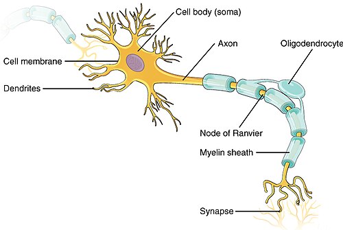
脱分極は、人体内の多くの細胞の機能にとって不可欠であり、ニューロン内およびニューロン間の刺激伝達はその典型例である。刺激の受容、それらの刺激の神経統合、そして刺激に対するニューロンの応答はすべて、ニューロンが脱分極を利用してニューロン内またはニューロン間で刺激を伝達する能力に依存している。
ニューロンへの刺激は、物理的、電気的、または化学的なものであり、刺激されたニューロンを抑制することも興奮させることもできます。抑制刺激はニューロンの樹状突起に伝達され、ニューロンの過分極を引き起こします。抑制刺激後の過分極は、ニューロン内の電圧を静止電位よりもさらに低下させます。抑制刺激によってニューロンが過分極すると、脱分極が起こるためには、より大きな負電荷を克服する必要があります。
一方、興奮刺激はニューロン内の電圧を上昇させ、その結果、静止状態のニューロンよりも脱分極しやすくなる。興奮性刺激であろうと抑制性刺激であろうと、刺激はニューロンの樹状突起を通って細胞体へと伝わり、そこで統合される。

刺激が細胞体に到達すると、神経は応答する前に様々な刺激を統合する必要があります。樹状突起を伝わってきた刺激は軸索丘で収束し、そこで合計されて神経細胞の応答が決定されます。刺激の合計が閾値電位と呼ばれる一定の電圧に達すると、脱分極が軸索丘から軸索へと続きます。
The surge of depolarization traveling from the axon hillock to the axon terminal is known as an action potential. Action potentials reach the axon terminal, where the action potential triggers the release of neurotransmitters from the neuron. The neurotransmitters that are released from the axon continue on to stimulate other cells such as other neurons or muscle cells. After an action potential travels down the axon of a neuron, the resting membrane potential of the axon must be restored before another action potential can travel the axon. This is known as the recovery period of the neuron, during which the neuron cannot transmit another action potential.
The importance and versatility of depolarization within cells can be seen in the relationship between rod cells in the eye and their associated neurons. When rod cells are in the dark, they are depolarized. In the rod cells, this depolarization is maintained by ion channels that remain open due to the higher voltage of the rod cell in the depolarized state. The ion channels allow calcium and sodium to pass freely into the cell, maintaining the depolarized state. Rod cells in the depolarized state constantly release neurotransmitters which in turn stimulate the nerves associated with rod cells. This cycle is broken when rod cells are exposed to light; the absorption of light by the rod cell causes the channels that had facilitated the entry of sodium and calcium into the rod cell to close. When these channels close, the rod cells produce fewer neurotransmitters, which is perceived by the brain as an increase in light. Therefore, in the case of rod cells and their associated neurons, depolarization actually prevents a signal from reaching the brain as opposed to stimulating the transmission of the signal.[15]

Depolarization occurs in the four chambers of the heart: both atria first, and then both ventricles.
This process continues regularly, unless there is a problem in the heart.[16]
In a normal ECG:[17]
There are drugs, called depolarization blocking agents, that cause prolonged depolarization by opening channels responsible for depolarization and not allowing them to close, preventing repolarization. Examples include the nicotinic agonists, suxamethonium and decamethonium.[18] Succinylcholine (suxamethonium) activates nicotinic acetylcholine receptors and keeps the endplate depolarized. It functionally blocks transmission because voltage-gated sodium channels remain inactivated.[19] Decamethonium is an older depolarizing blocker with a similar principle and of interest for many researchers.[20]