Loading article…
一対の顎下腺(歴史的には顎下腺として知られている)は、口底の下に位置する主要な唾液腺である。成人では、それぞれ約15グラムの重さがあり、刺激を受けていない唾液分泌の約60~67%を占める。刺激を受けると、耳下腺の分泌が50%まで増加するにつれて、顎下腺の寄与は比例して減少する。[ 1 ]正常な成人の顎下腺の平均長さは約27mm 、平均幅は約14.3mmである。[ 2 ]

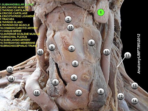
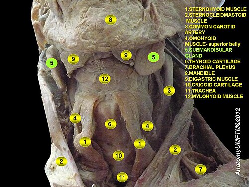
各顎下腺は表層部と深層部に分かれており、これらは顎舌骨筋によって隔てられている。[ 3 ]
顎下腺管(ワルトン管とも呼ばれる)は、 長さ約5cmである。腺の深部から始まり、口腔底を前方に走る。舌神経は管の下をループ状に回り、管の外側から内側へと通過する。管は舌小帯の両側にある舌下小丘に開口する。[ 4 ]
頭を前方に傾けた状態で、下顎角付近の下縁から内側に移動させることで、下顎体の下方後方に両側から腺を触診することができる。 [ 5 ]
顎下腺管の末端部は口腔底に位置し、顎下腺管乳頭の開口部を通って開口する。管の位置と0.5~1.5mm 幅の開口部の位置は常に左右対称だが、予測不可能である。そのため、顎下腺管乳頭の認識は時に困難となることがある。Aničinらは、乳頭の肉眼的外観と唾液腺内視鏡アプローチに基づいて、顎下腺乳頭をA型、B型、C型、D型の4種類に分類した。[ 6 ]
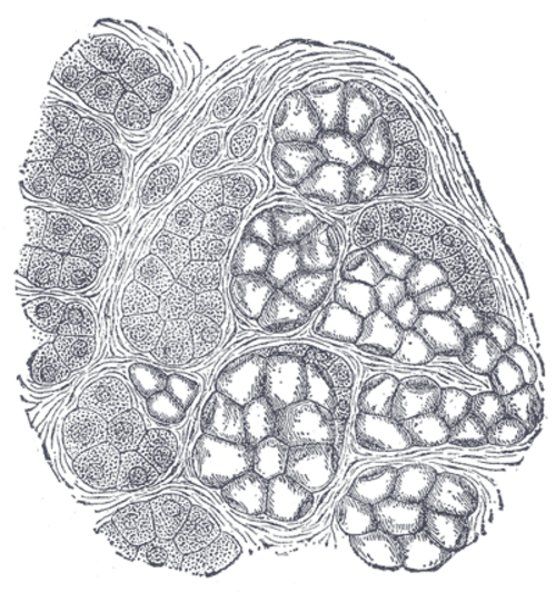
葉はさらに小さな小葉からなり、小葉には腺の分泌単位である腺小体が含まれています。各腺小体には1つ以上の腺房(または腺胞)が含まれており、腺房は分泌物を導管に分泌する小さな細胞の集まりです。
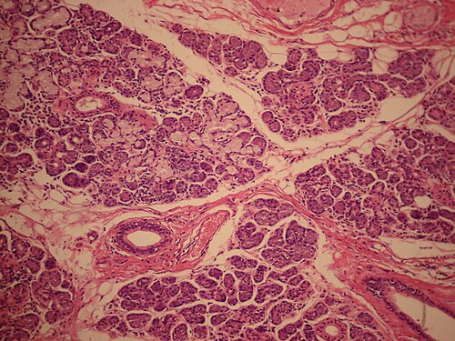
顎下腺は、漿液性分泌細胞と粘液性分泌細胞の両方を含む混合型の分岐管状腺房である。漿液性腺房が優勢であり、粘液性腺房は分泌単位のごく一部を占める。[ 7 ]
漿液細胞は、唾液アミラーゼなどの酵素を含む、水様でタンパク質が豊富な分泌物を産生する。粘液細胞は、口腔内や食物塊を潤滑するムチンを産生する。一部の粘液腺房は、三日月形の漿液性半月状構造で覆われているように見えることがある。
主腺房分泌物は導管系を通過する際に変化する。この腺には比較的短い介在導管と発達した線条導管があり、これらが唾液の電解質組成を変化させる。[ 7 ]
この腺は顔面動脈と舌動脈の枝から動脈血供給を受けています。[ 8 ]特に顎下動脈と舌下動脈から血液供給を受け、顔面静脈と舌静脈から血液が排出されます。
顎下腺のリンパ管は、まず顎下リンパ節に流れ込み、その後、頸静脈二腹筋リンパ節を含む上部深頸リンパ節に流れ込む。
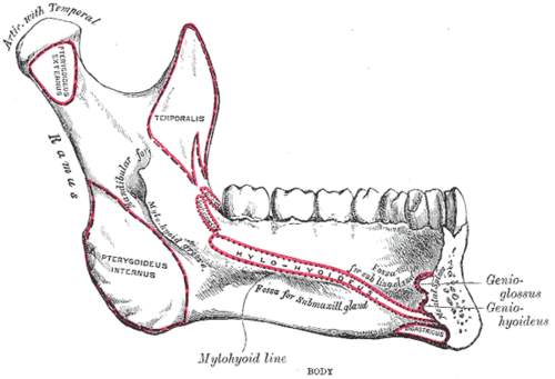
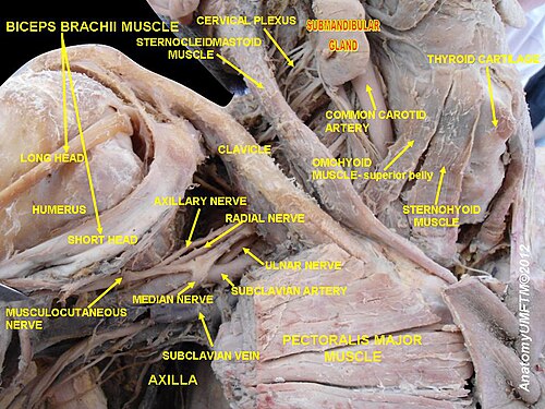
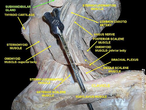
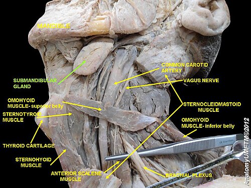
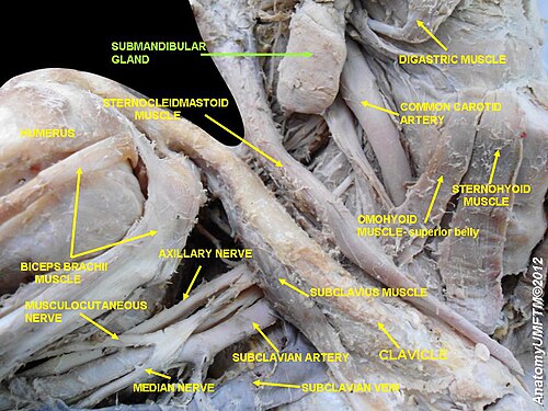
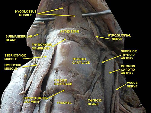
The submandibular gland occupies most of the submandibular triangle and rests partly within the submandibular fossa on the medial surface of the mandible. Its larger superficial part lies inferior to the mylohyoid muscle, while its smaller deep part curves around the posterior border of the mylohyoid and enters the floor of the mouth.[9]
The facial artery has a close relationship with the posterior and deep surfaces of the gland, while the facial vein generally passes superficially. The marginal mandibular branch of the facial nerve lies superficial to the facial vessels and is an important surgical relation.[9]
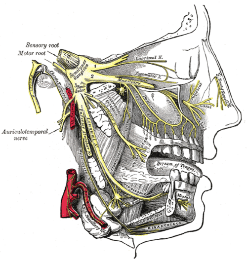
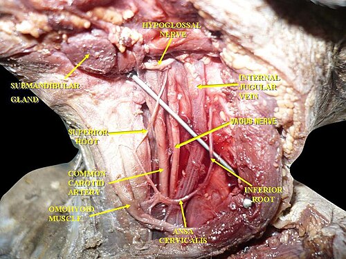
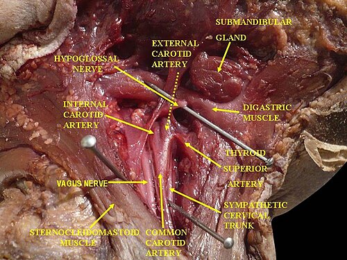
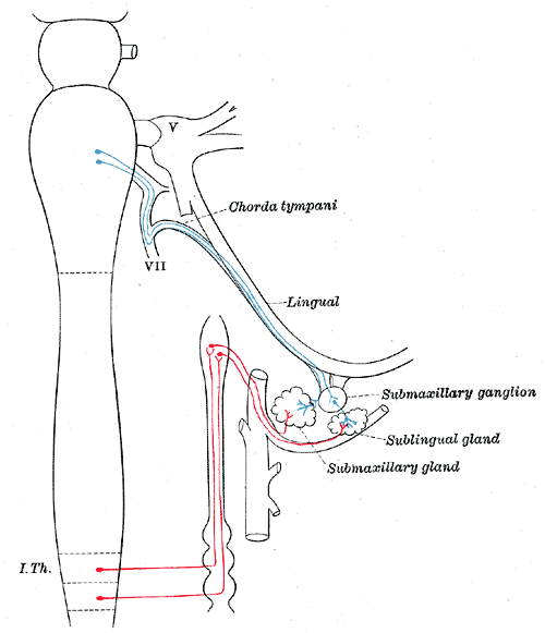
The lingual nerve and submandibular ganglion are closely related to the deep part of the gland and the submandibular duct. The lingual nerve passes beneath the duct as it travels from the lateral to the medial side. The hypoglossal nerve lies deep and inferior to the duct and gland.[9]
The submandibular salivary glands develop later than the parotid glands and appear late in the sixth week of prenatal development. They develop bilaterally from epithelial buds in the sulcus surrounding the sublingual folds on the floor of the primitive mouth. Solid cords branch from the buds and grow posteriorly, lateral to the developing tongue.
The cords of the submandibular gland later branch further and become canalized to form the ductal system. The submandibular gland acini develop from the rounded terminal ends of the cords at approximately 12 weeks, and secretory activity through the submandibular duct begins at approximately 16 weeks.
Growth of the submandibular gland continues after birth through the formation of additional acini. Lateral to both sides of the tongue, a linear groove develops and closes to form the submandibular duct.[10]
The submandibular glands are one of the three paired major salivary glands. The other two are the parotid and sublingual glands.
The submandibular glands produce most of the saliva secreted under unstimulated resting conditions. During stimulation, such as chewing, the relative contribution of the parotid glands increases.[4]
Their mixed serous and mucous secretion lubricates the oral mucosa and food bolus, assists mastication, swallowing and speech, and begins starch digestion through the activity of salivary amylase.[7]
Saliva also contributes to oral protection. Mucins help lubricate oral tissues, bicarbonate helps buffer acids, and components including immunoglobulins, lysozyme and lactoferrin contribute to antimicrobial defence.[7]

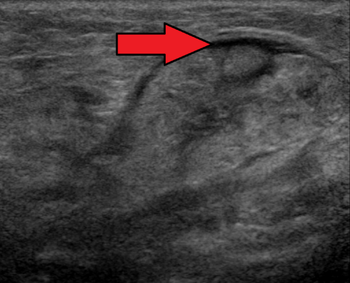
The submandibular gland is commonly affected by salivary duct calculi, also known as sialoliths. Its susceptibility has been associated with the long, upward course of the submandibular duct and the character of its mixed secretion.[9]
Obstruction of the duct may cause recurrent pain and swelling, particularly during meals. Persistent obstruction can promote salivary stasis, inflammation and secondary bacterial sialadenitis.[9]
Small or accessible calculi may be treated using gland massage, duct dilation, transoral removal, lithotripsy or sialoendoscopy. Recurrent obstruction, chronic sialadenitis or neoplasia may require excision of the gland.[9]
Structures at risk during submandibular gland surgery include the marginal mandibular branch of the facial nerve, the lingual nerve, the hypoglossal nerve, and the facial artery and vein.[9]
Benign and malignant tumours can also develop in the submandibular gland.