
Loading article…
Geniom RT Analyzerは、分子生物学における診断検査に使用される装置です。Geniom RT Analyzerは、組織マイクロRNAレベルの動的な性質を疾患進行のバイオマーカーとして利用します。Geniomアナライザーは、マイクロ流体プライマー伸長アッセイ(MPEA)技術(図2)を介してマイクロRNAを定量するために、マイクロ流体およびバイオチップマイクロアレイ技術を組み込んでいます。[ 1 ]
多くのヒト疾患(例えば癌)は、分子レベルで動的なマイクロRNAレベルに関与していると考えられている。マイクロRNAは遺伝子発現の調節に重要であり、したがって癌遺伝子の活性化や腫瘍抑制遺伝子の不活性化に重要な影響を与える可能性がある。[ 2 ]特定の種類の癌の進行全体を通して、特定のマイクロRNAが異なるレベルで検出されている。[ 3 ]
早期診断検査は、多くのヒト疾患において課題であることが証明されています[ 4 ]。症状表現型は曖昧であったり、微妙であったりする可能性があるためです。その結果、分子バイオマーカーの特性評価に特化した広範な研究分野が発展しました。バイオマーカーは、さまざまな疾患段階における生物学的分子の正確な測定を可能にします。これらの測定は、将来の早期診断検査に役立つ豊富なデータソースに最終的に貢献します。Geniom RT Analyzerは、マイクロRNAバイオマーカーの自動プロファイリングを実行し、この増大するデータソースに貢献します[ 5 ] 。
マイクロRNAレベルの検出は、空間的、時間的、あるいはその両方を組み合わせた場合において有用である。
空間的マイクロRNAダイナミクス: 最近の発見により、血液中に遊離したマイクロRNAが検出されました。[ 2 ]これらの遊離したマイクロRNAは、組織の細胞内のマイクロRNAと比較して、内因性RNAase活性から保護されていることがわかっています。この保護により、これらの遊離したマイクロRNAは適切な安定したバイオマーカーとなります。この安定したバイオマーカーは、組織間でマイクロRNAレベルを比較する際のコントロールとして使用できます。マイクロRNAレベルを比較し、組織間の発現パターンを評価することは、癌や疾患の分類など、さまざまな用途に使用できます。[ 6 ]
Temporal microRNA dynamics: A complication with designing and developing tumour suppressor drugs for cancer patients is that the molecular environment within and around the tumour does not seem to be constant throughout the process of tumour development.[7] MicroRNAs are one class of molecules that contribute to this environmental fluctuation for the tumour. Detecting microRNA levels in tumour or regular tissues over time can help to gain insight on the nature of these environmental changes. This information can in turn be used in deciding when the proper timing is for various therapeutic interventions.
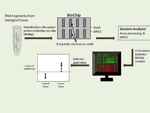
The platform for the Geniom RT Analyzer consists of a biochip containing 8 separate microarrays. Tissue derived microRNAs do not require treatment prior to introduction to the biochip. The biochip contains validated and optimized customizable capture probes. The on-chip Microfluidic Primer Extension Assay (MPEA) enables direct microRNA-capture probe hybridization. After biotin labelling, primer extension and washing, the Geniom Analyzer undergoes automated processing of the arrays. A charge-coupled device (CCD) camera assists the biochip readout, which displays a pictorial image of the microRNA quantification. This image is supported by the use of biotinylated nucleotides and subsequent staining with a streptavidin-phycoerythrin conjugate. Due to the use of 8 separate microarrays per biochip, seven replicate intensity readings are made available and the median value is generally applied to the graphical results. An analysis of biomarker assessment is then made and data can be stored in the miRDBXP database.
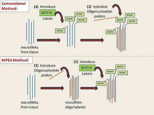
MPEA: The MPEA technique utilizes microfluidic technology to ensure the correct timing and, consequently, the correct alignment of the capture probes with the microRNA molecules. Conventional methods of probe-microRNA hybridization require prior treatment of microRNAs with potentially costly reagents. These conventional methods also require initial introduction of biotin prior to hybridization. Due to the application of microfluidics, neither initial biotin nor reagent treatment is necessary prior to primer extension. Rather, the unlabeled, hybridized microRNA behaves as the primer for enzymatic elongation, a process in which biotinylated nucleotides are assembled.