
Loading article…
ソノマイクロメトリーは、圧電結晶が埋め込まれた媒体中の音響信号の速度に基づいて、圧電結晶間の距離を測定する技術です。通常、結晶はエポキシ樹脂製の「レンズ」でコーティングされ、互いに向き合うように媒体中に配置されます。いずれかの結晶に送られた電気信号は音に変換され、媒体中を伝わり、最終的にもう一方の結晶に到達します。そこで音は電気信号に変換され、受信機によって検出されます。結晶間を音が伝わるのにかかる時間と媒体中の音速から、結晶間の距離を計算することができます。
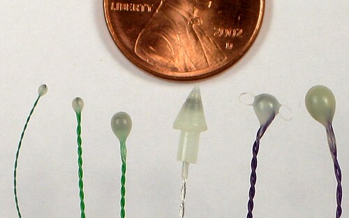
超音波マイクロメトリーは、1956年にディーン・フランクリンによって実験動物の心臓機能の研究に初めて応用され、その後、生体力学を研究する生物学者をはじめ、他の生理学的器官系や構造(消化器系、泌尿生殖器系、筋骨格系)の研究にも急速に採用されました。医療機器メーカーも、開発の研究開発段階において、機器の物理的性能、耐久性、寿命を評価するために超音波マイクロメトリーを使用しています。現在、超音波マイクロメトリーは、動物の運動、摂食、その他の生体力学的機能における筋肉の長さの変化を測定する最も一般的な方法となっています。
数十年前、最初に開発された当時は、結晶間の信号検出が適切に行われるよう、結晶の向きを正しく調整することに細心の注意が払われていたが、より現代的なソノマイクロメーターのハードウェア(一般的には1995年以降に開発されたもの)では、結晶の向きにそのような注意を払う必要はない。
超音波マイクロメーターシステムは、科学者、技術者、生物学者によって、軟質固体、ゲル、流体の物性を測定するために使用されます。筋肉組織の力学的特性や機能を測定するために使用されていますが、これらの装置に医学的または治療的な用途は知られていません。超音波マイクロメーター装置は、動物や人間の病状の治療や診断には使用されません。
ソノマイクロメーターによる測定を行う際の主要な基準は、結晶間の空間または物質が、100 kHz から少なくとも数 MHz の周波数範囲で超音波伝導能力を有することです。これは通常、結晶が流体、ゲル、軟質固体、および一部の硬質固体からなる媒体内または媒体上で動作する必要があることを意味し、これには生体組織(血液、筋肉、脂肪など)も含まれます。結晶間に空気の隙間や固体物体が存在すると、結晶間の距離の測定が困難になるか、完全に不可能になります。
Sonomicrometer measurements are only accurate when the medium surrounding the crystals or the object the crystals are measuring has a uniform speed of sound. In biological settings, almost all tissues have a speed of sound in the range of 1550 to 1600 meters per second, which is satisfactorily uniform to give accurate measurements to within 3% to 4%, and more typically to within 1%.
A sonomicrometer device measures distance by performing a timing measurement of the sound transmitted by one crystal and received by one (or more) adjacent crystals. The ability to resolve small differences in this transit-time (or "time-of-flight") directly correlates with the ability to resolve small changes in the distances between crystals. The first generation of sonomicrometer devices were analog devices that integrated the time-of-flight as a voltage ramp function. Their resolution was a function of the slope of the ramp and the electrical noise of system components. Those systems output the length measurements as an analog voltage. The second generation of sonomicrometer devices were digital—they measure time-of-flight by incrementing high-speed digital counters. Typically these are 12 to 16-bit counters operating from 32 to 128 MHz. A digital system will output length measurements in form of a digital number (the time-of-flight count value). Because of their digital nature, the resolution can be exactly specified as a function of counter clock frequency. For example, when making measurements in biological tissue at body temperature (velocity = 1,590 m/s), a digital sonomicrometer operating with a clock speed of 128 MHz will have a spatial resolution of 12 micrometres.
Sonomicrometry is often used in studies of animal physiology where precise distances at high temporal resolution are needed, particularly when such distances are not externally measurable. Sonomicrometry crystals are most commonly implanted within skeletal or cardiac muscle tissue to track length changes during an activity (heartbeat, flapping a wing, chewing, etc.). However, they can be very useful for tracking movement of entire structures which are not visible but immersed in fluid, such as the bones in the mouth of a fish during feeding.